Fraud Blocker Skip to main content

Fluorescence Micrograph Collection

"Unlocking the Hidden World

Background imageFluorescence Micrograph Collection: Nerve and glial cells, light micrograph

Nerve and glial cells, light micrograph
Nerve and glial cells, fluorescence light micrograph. These are neural stem cells that have differentiated into neurons (nerve cells, blue) and glial cells (support cells, red)

Background imageFluorescence Micrograph Collection: Dividing cells

Dividing cells. Computer artwork of a spiral of cells during mitosis (nuclear division). During mitosis two daughter nuclei are formed from one parent nucleus

Background imageFluorescence Micrograph Collection: Embryonic smooth muscle cell C018 / 8595

Embryonic smooth muscle cell C018 / 8595
Embryonic smooth muscle cell, immunofluorescence micrograph. Actin filaments are green, the protein vinculin is light blue. Actin is part of the cytoskeleton

Background imageFluorescence Micrograph Collection: Neural progenitor cell differentiation C018 / 8758

Neural progenitor cell differentiation C018 / 8758
Neural progenitor cell differentiation. Fluorescence light micrograph of neural progenitor cells that have been grown in a medium that is selective for astrocytes for three weeks

Background imageFluorescence Micrograph Collection: Actin-based motility, light micrograph C017 / 8333

Actin-based motility, light micrograph C017 / 8333
Actin-based motility. 3D model made from optical sections of Shigella sp. bacteria (pink) infecting cells. Filaments of the protein actin, which is part of the cells cystoskeleton

Background imageFluorescence Micrograph Collection: Mitosis, fluorescence micrograph

Mitosis, fluorescence micrograph
Mitosis. Fluorescence micrograph of a cell (centre) in anaphase during mitosis (nuclear division). During mitosis two daughter nuclei are formed from one parent nucleus

Background imageFluorescence Micrograph Collection: Cytokinesis

Cytokinesis. Fluorescent micrograph of an animal cell during cytokinesis (cell division). Cytokinesis occurs after nuclear division (mitosis), which produces two daughter nuclei

Background imageFluorescence Micrograph Collection: Abnormal mitosis

Abnormal mitosis
Mitosis. Fluorescence micrograph of a cell during abnormal anaphase of mitosis (nuclear division). During mitosis two daughter nuclei are formed from one parent nucleus

Background imageFluorescence Micrograph Collection: Cell division

Cell division. Fluorescent micrograph of an animal cell during cytokinesis (cell division). Cytokinesis occurs after nuclear division (mitosis)

Background imageFluorescence Micrograph Collection: Mitosis

Mitosis. Fluorescence micrograph of a cell during prometaphase of mitosis (nuclear division). During mitosis two daughter nuclei are formed from one parent nucleus

Background imageFluorescence Micrograph Collection: Macrophages and tuberculosis vaccine

Macrophages and tuberculosis vaccine. Fluorescence light micrograph of macrophage (red, outlined in black) white blood cells that have phagocytosed (engulfed) Mycobacterium bovis bacteria (pink)

Background imageFluorescence Micrograph Collection: DNA

DNA
Deoxyribonucleic acid. Conceptual artwork of DNA (deoxyribonucleic acid) spelt out with fluoresence micrographs of anaphase stage cells. Anaphase is a stage of mitosis (nuclear division)

Background imageFluorescence Micrograph Collection: Stentor ciliate protozoa light micrograph

Stentor ciliate protozoa light micrograph

Background imageFluorescence Micrograph Collection: Thale cress anther and pollen, micrograph

Thale cress anther and pollen, micrograph
Thale cress anther and pollenr. Fluorescence micrograph of an anther (male reproductive structure) from a thale cress (Arabidopsis thaliana) flower, showing the pollen grains (round) inside

Background imageFluorescence Micrograph Collection: Thale cress stamens, micrograph

Thale cress stamens, micrograph
Mitosis. Confocal light micrograph of the stages mitosis (nuclear division) and cytokinesis (cell division). During mitosis the nuclear envelope disintegrates (3rd image) and the chromosomes (purple)

Background imageFluorescence Micrograph Collection: Thale cress flower, micrograph

Thale cress flower, micrograph
Thale cress flower. Fluorescence micrograph of a flower from a thale cress (Arabidopsis thaliana) plant. The female reproductive structures - carpel (red) and stigma (yellow) - can be seen at centre

Background imageFluorescence Micrograph Collection: Hippocampal neuron fluorescent micrograph

Hippocampal neuron fluorescent micrograph
Hippocampal neuron. Fluorescent micrograph of a neuron (nerve cell, centre) from the hippocampus, surrounded by glial cells (support cells)

Background imageFluorescence Micrograph Collection: Leaf hairs, fluorescence micrograph

Leaf hairs, fluorescence micrograph
Leaf hairs. Fluorescence micrograph showing leaf hairs (trichomes) on a Nicotiana benthamiana plant. A fluorescence microscope is an optical microscope that uses fluorescence

Background imageFluorescence Micrograph Collection: Thale cress anther, micrograph

Thale cress anther, micrograph
Thale cress anther. Fluorescence micrograph of an anther (male reproductive structure) from a thale cress (Arabidopsis thaliana) flower, showing the pollen grains (red, round) inside

Background imageFluorescence Micrograph Collection: Thale cress leaf hair, micrograph

Thale cress leaf hair, micrograph
Thale cress leaf hair. Fluorescence micrograph showing the microfilaments of a leaf hair (trichome) from a thale cress (Arabidopsis thaliana) plant

Background imageFluorescence Micrograph Collection: Cell structure, fluorescent micrograph

Cell structure, fluorescent micrograph
Cell structure. Fluorescent light micrograph of cultured cells from a cell line derived from African green monkey kidney cells. Microtubules, part of the cells cytoskeleton, are red

Background imageFluorescence Micrograph Collection: Thale cress stigma, micrograph

Thale cress stigma, micrograph
Thale cress stigma. Fluorescence micrograph of a stigma (female reproductive structure) from a thale cress (Arabidopsis thaliana) flower

Background imageFluorescence Micrograph Collection: Stamens and carpels, micrograph

Stamens and carpels, micrograph
Stamens and carpels. Fluorescence micrograph of stamens (male reproductive structures, far left) and carpels (female reproductive structures)

Background imageFluorescence Micrograph Collection: Thale cress stigma, micrograph

Thale cress stigma, micrograph
Thale cress stigma. Fluorescence micrograph of a stigma (female reproductive structure) from a thale cress (Arabidopsis thaliana) flower

Background imageFluorescence Micrograph Collection: Caenorhabditis elegans, micrograph

Caenorhabditis elegans, micrograph
Caenorhabditis elegans. Fluorescence micrograph of the transparent nematode (roundworm) worm Caenorhabditis elegans. A fluorescence microscope is an optical microscope that uses fluorescence

Background imageFluorescence Micrograph Collection: Keratinocyte skin cells, light micrograph

Keratinocyte skin cells, light micrograph
Keratinocyte skin cells. Fluorescent light micrograph of the cytoskeleton of human keratinocyte skin cells. Cell nuclei are oval. The rest of the cells contents have been biochemically extracted


All Professionally Made to Order for Quick Shipping

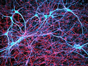
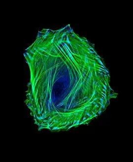
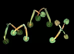
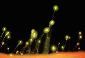
"Unlocking the Hidden World: Exploring Fluorescence Micrographs" Step into a mesmerizing realm where nerve and glial cells reveal their intricate beauty under the lens of a fluorescence micrograph. This captivating light micrograph unveils the delicate network of these vital components, showcasing their role in transmitting signals throughout our bodies. Witness the awe-inspiring process of cell division as dividing cells gracefully dance across this microscopic stage. Each moment captured in this fluorescent masterpiece showcases the remarkable complexity and precision involved in creating new life. Delve deeper into nature's wonders with a glimpse at a Thale cress flower through the lens of a micrograph. The vibrant hues and intricate patterns highlight its exquisite structure, reminding us of the astonishing diversity found within even the tiniest organisms. Embark on an exploration of embryonic smooth muscle cells, as they take center stage in another stunning fluorescence micrograph. Witness their development unfold before your eyes, capturing every step towards becoming fully functional muscles that power our movements. Journey alongside neural progenitor cells as they undergo differentiation, showcased vividly in yet another breathtaking fluorescence micrograph. Marvel at how these specialized cells transform into various types within our complex nervous system, paving the way for proper brain function. Behold an actin-based motility phenomenon captured through a light micrograph – an enchanting display of cellular movement orchestrated by this essential protein. Witness its crucial role in shaping cellular architecture and facilitating numerous biological processes. The symphony continues with multiple glimpses into mitosis - nature's choreographed spectacle of cell division depicted repeatedly throughout this collection. Observe each meticulous phase as chromosomes align, duplicate, separate, ensuring accurate distribution to daughter cells during this fundamental process. However, amidst harmony lies occasional discordance; witness abnormal mitosis caught frozen in time within one striking image. Explore anomalies that challenge normalcy but provide valuable insights into understanding diseases such as cancer or genetic disorders affecting cell division dynamics.