
Metatarsal Collection
The metatarsal, a vital component of the human foot, plays a crucial role in maintaining balance and facilitating movement


All Professionally Made to Order for Quick Shipping
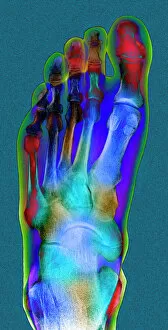
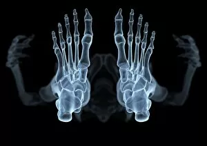
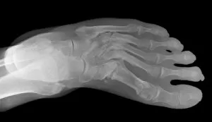
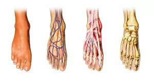
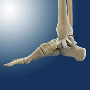
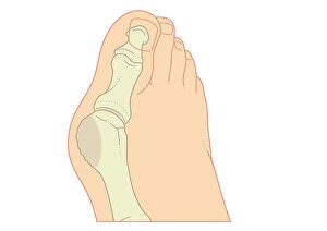
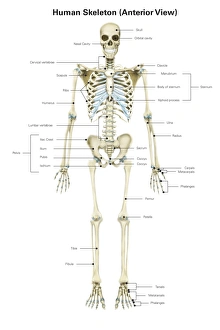
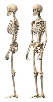
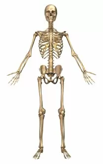
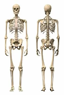
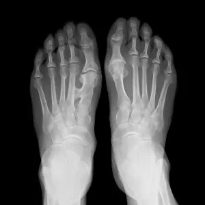
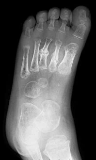
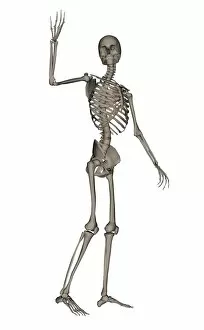
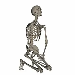
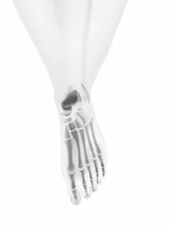
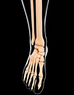
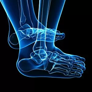
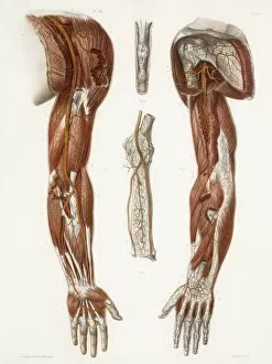
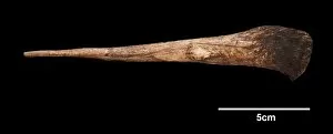
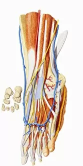
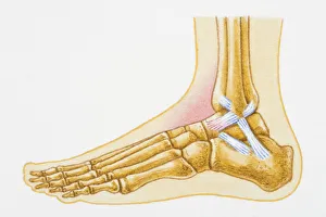
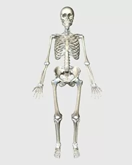
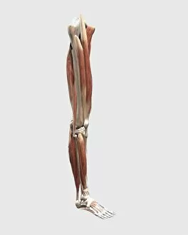
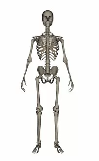
The metatarsal, a vital component of the human foot, plays a crucial role in maintaining balance and facilitating movement. In its normal state, it forms part of the intricate skeletal structure that supports our body weight. When viewed through an X-ray, this bone reveals its elegant design and alignment within the foot. However, not all metatarsals tell the same story. A Lisfranc fracture can disrupt this harmony, causing immense pain and hindering mobility. An X-ray artwork vividly captures this injury's impact on the delicate bones. Exploring further back in time, we encounter fascinating glimpses into our evolutionary history. The cast of an Australopithecine or Homo habilis foot (OH8) showcases how our ancestors' feet differed from ours today. Moving to another angle, inner ankle ligaments come into focus through captivating artwork C013/4451. These ligaments provide stability to our ankles during various activities. Zooming out to encompass the entire skeletal system brings forth a comprehensive understanding of how each bone interacts with others for optimal functionality. From front view to anterior view with labels, we unravel the complexity behind our physical framework. Not limited to humans alone, even animals have left their mark on metatarsals throughout history. An intricately engraved horse-head serves as a testament to ancient artistry etched onto these bones. Returning to present-day anatomy, dynamic postures showcase how male human skeletons adapt and respond during movement. Both front and side views offer unique perspectives on their form and function.