SARS coronavirus, artwork C016 / 3053

![]()
Wall Art and Photo Gifts from Science Photo Library
SARS coronavirus, artwork C016 / 3053
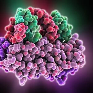
SARS coronavirus proteins. Cutaway computer model showing the protein structure of a SARS coronavirus particle (virion). SARS (severe acute respiratory syndrome) is a viral respiratory illness caused by the SARS-associated coronavirus (SARS-CoV); a single stranded RNA (ribonucleic acid, blue coil) virus. Each SARS-CoV virion is made up of four major structural proteins: nucleocapsid, spike trimer (red), membrane (yellow) and envelope (purple), plus HE haemagglutinin-esterase dimer (orange)
Science Photo Library features Science and Medical images including photos and illustrations
Media ID 9202855
© RAMON ANDRADE 3DCIENCIA/SCIENCE PHOTO LIBRARY
Biomolecule Coronavirus Cutaway Dimer Envelope Genetic Haemagglutinin Hemagglutinin Inside Internal Macromolecule Macromolecules Membrane Micro Organism Micro Organisms Microbiology Microorganism Microorganisms Molecular Biology Particle Pentamer Proteins Proteomics Ribonucleic Acid Rna Virus Sars Severe Acute Respiratory Syndrome Single Strand Single Stranded Spike Structural Trimer Viral Virion Virological Virology Biochemical Biochemistry Cutouts Genetics Microbiological Molecular Model Molecular Structure Virus
EDITORS COMMENTS
This print showcases the intricate protein structure of a SARS coronavirus particle, offering a glimpse into the world of virology and molecular biology. Created by RAMON ANDRADE 3DCIENCIA/SCIENCE PHOTO LIBRARY, this artwork titled "SARS coronavirus, artwork C016 / 3053" presents a cutaway computer model that reveals the internal composition of the virion. The SARS-associated coronavirus (SARS-CoV) is responsible for causing severe acute respiratory syndrome (SARS), a viral respiratory illness. The single-stranded RNA virus is depicted as a blue coil in this image, highlighting its genetic nature. Comprising four major structural proteins - nucleocapsid, spike trimer (in striking red), membrane (in vibrant yellow), and envelope (in regal purple) - along with HE haemagglutinin-esterase dimer in orange coloration. With its white background and precise detailing, this illustration allows us to appreciate the complexity and beauty found within these microscopic entities. It serves as an invaluable tool for researchers studying SARS-CoV and aids in understanding how it interacts with human cells during infection. Through biomolecular analysis and proteomics research, scientists can unravel crucial information about viruses like SARS-CoV. This visual representation provides insights into their macromolecular structures while shedding light on potential targets for therapeutic interventions. RAMON ANDRADE's creation not only captures the essence of scientific exploration but also reminds us of our ongoing battle against infectious diseases. As we delve deeper into understanding these microorganisms at their molecular level, we inch closer towards finding effective treatments or preventive measures to combat them effectively.
MADE IN THE UK
Safe Shipping with 30 Day Money Back Guarantee
FREE PERSONALISATION*
We are proud to offer a range of customisation features including Personalised Captions, Color Filters and Picture Zoom Tools
SECURE PAYMENTS
We happily accept a wide range of payment options so you can pay for the things you need in the way that is most convenient for you
* Options may vary by product and licensing agreement. Zoomed Pictures can be adjusted in the Basket.