Fraud Blocker Skip to main content

Ribonucleic Acid Collection

"Unraveling the Secrets of Ribonucleic Acid: The Double-Stranded RNA Molecule" In the intricate world of molecular biology

Background imageRibonucleic Acid Collection: DNA transcription, molecular model

DNA transcription, molecular model. Secondary structure of the enzyme RNA polymerase II synthesising a mRNA (messenger ribonucleic acid, lilac) strand from a DNA (deoxyribonucleic acid)

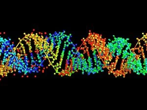
Background imageRibonucleic Acid Collection: Double-stranded RNA molecule

Double-stranded RNA molecule. Computer model of the structure of double-stranded RNA (ribonucleic acid). The majority of RNA in a cell is in the single-stranded form

Background imageRibonucleic Acid Collection: Bacterial ribosome

Bacterial ribosome. Computer model showing the secondary structure of a 30S (small) ribosomal sub-unit from the bacteria Thermus thermophilus

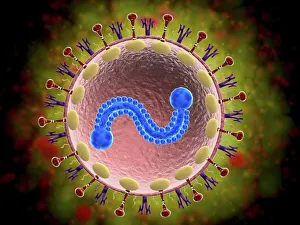
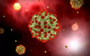
Background imageRibonucleic Acid Collection: Microscopic view of human respiratory syncytial virus

Microscopic view of human respiratory syncytial virus (RSV). RSV causes respiratory tract infection of the lungs and breathing passages

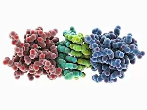
Background imageRibonucleic Acid Collection: RNA-editing enzyme, molecular model

RNA-editing enzyme, molecular model
RNA-editing enzyme. Molecular model of a left-handed, RNA double helix (Z-RNA, centre) bound by the Z alpha domain of the human RNA-editing enzyme ADAR1 (double-stranded RNA adenosine deaminase)

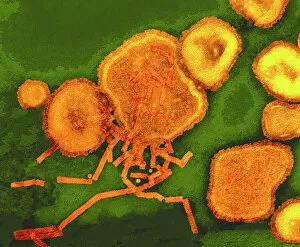
Background imageRibonucleic Acid Collection: Paramyxovirus particles, TEM

Paramyxovirus particles, TEM
Sendai virus. Coloured transmission electron micrograph (TEM) of Sendai virus particles (virions, orange). The protein coat (capsid) of one of the particles has split

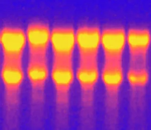
Background imageRibonucleic Acid Collection: Electrophoresis of RNA

Electrophoresis of RNA
Liver RNA. Electrophoresis gel containing RNA (ribonucleic acid) isolated from liver tissue. The RNA molecules (white bands) are being observed under ultraviolet light

Background imageRibonucleic Acid Collection: Stylized rabies virus particles

Stylized rabies virus particles, the cause of the viral neuroinvasive disease acute encephalitis

Background imageRibonucleic Acid Collection: Microscopic view of yellow fever virus

Microscopic view of yellow fever virus. Yellow fever is an acute viral disease

Background imageRibonucleic Acid Collection: Conceptual image of rabies virus

Conceptual image of rabies virus

Background imageRibonucleic Acid Collection: Argonaute protein and microRNA F006 / 9752

Argonaute protein and microRNA F006 / 9752
Argonaute protein. Molecular model of human argonaute-2 protein complexed with microRNA (micro ribonucleic acid). This protein is part of the RNA-induced silencing complex (RISC)

Background imageRibonucleic Acid Collection: RNA-induced silencing complex F006 / 9586

RNA-induced silencing complex F006 / 9586
RNA-induced silencing complex (RISC), molecular model. This complex consists of a bacterial argonaute protein (top) bound to a small interfering RNA (siRNA) molecule (red and blue)

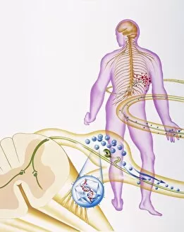
Background imageRibonucleic Acid Collection: Shingles nerve damage

Shingles nerve damage

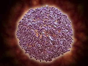
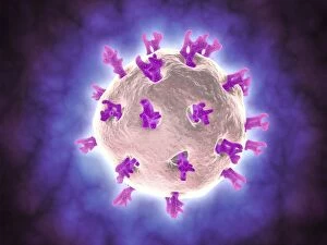
Background imageRibonucleic Acid Collection: Single virus particle

Single virus particle

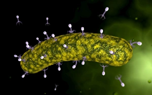
Background imageRibonucleic Acid Collection: Microscopic view of bacteriophages on the surface of a bacteria

Microscopic view of bacteriophages on the surface of a bacteria

Background imageRibonucleic Acid Collection: Conceptual image of RNA virus replication

Conceptual image of RNA virus replication

Background imageRibonucleic Acid Collection: Cluster of HIV virus

Cluster of HIV virus. HIV is the human immunodeficiency virus that can lead to acquired immune deficiency syndrom, or AIDS

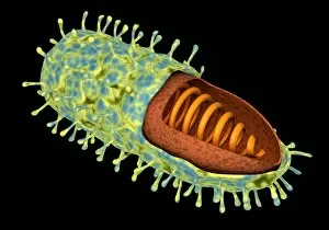
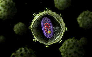
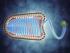
Background imageRibonucleic Acid Collection: Cutaway view of Reoviridae virus showing dna inside

Cutaway view of Reoviridae virus showing dna inside. Reoviruses can affect the gastronintestinal system and respiratory tract

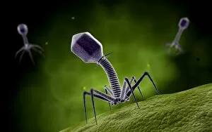
Background imageRibonucleic Acid Collection: Microscopic view of bacteriophage attacking bacteria

Microscopic view of bacteriophage attacking bacteria

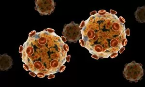
Background imageRibonucleic Acid Collection: Microscopic view of Sindbis virus

Microscopic view of Sindbis virus (SINV). SINV is a mosquito-borne virus that causes rash and arthritis, has been causing outbreaks in humans

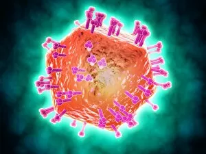
Background imageRibonucleic Acid Collection: Conceptual image of HIV virus

Conceptual image of HIV virus. HIV is the human immunodeficiency virus that can lead to acquired immune deficiency syndrome, or AIDS

Background imageRibonucleic Acid Collection: Microscopic view of HIV virus, cross section

Microscopic view of HIV virus, cross section

Background imageRibonucleic Acid Collection: Conceptual image of the Zika virus

Conceptual image of the Zika virus

Background imageRibonucleic Acid Collection: Microscopic view of respiratory syncytial virus

Microscopic view of respiratory syncytial virus (RSV). RSV is a common virus that leads to mild, cold-like symptoms in adults and children

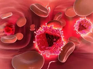
Background imageRibonucleic Acid Collection: Microscopic view of HIV virus inside the lungs

Microscopic view of HIV virus inside the lungs

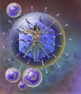
Background imageRibonucleic Acid Collection: Vitruvian Man inside virus particle

Vitruvian Man inside virus particle

Background imageRibonucleic Acid Collection: Microscopic view of bacteriophage

Microscopic view of bacteriophage

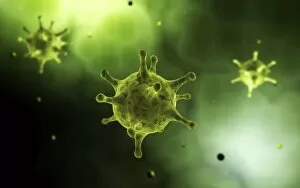
Background imageRibonucleic Acid Collection: Grouping of virus particles

Grouping of virus particles

Background imageRibonucleic Acid Collection: Microscopic view of virus

Microscopic view of virus

Background imageRibonucleic Acid Collection: Microscopic view of rotavirus

Microscopic view of rotavirus. Rotavirus is the most common cause of severe diarrhea among infants and young children. It is a genus of double-stranded RNA virus in the family Reoviridae

Background imageRibonucleic Acid Collection: Microscopic view of cell and virus

Microscopic view of cell and virus

Background imageRibonucleic Acid Collection: Microscopic view of Rubella virus

Microscopic view of Rubella virus
Microscopic view of Rubella. Rubella is an acute, contagious viral infection. While the illness is generally mild in children, it has serious consequences in pregnant women causing fetal death

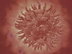
Background imageRibonucleic Acid Collection: Conceptual image of common virus

Conceptual image of common virus

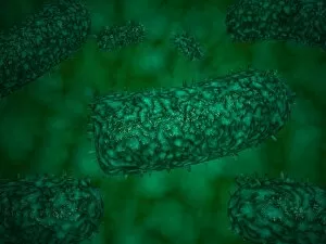
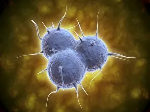
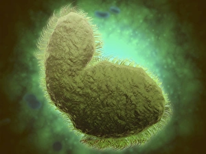
Background imageRibonucleic Acid Collection: Microscopic view of a microbe

Microscopic view of a microbe. Microbes are single-cell organisms so tiny that millions can fit into the eye of a needle

Background imageRibonucleic Acid Collection: Conceptual image of lyssavirus

Conceptual image of lyssavirus. Lyssavirus is a genus of viruses belonging to the family Rhabdoviridae. This group of RNA viruses includes the rabies virus traditionally associated with the disease

Background imageRibonucleic Acid Collection: Microscopic view of HIV virus

Microscopic view of HIV virus

Background imageRibonucleic Acid Collection: DNA transcription, illustration C018 / 0900

DNA transcription, illustration C018 / 0900
DNA (deoxyribonucleic acid) transcription. Illustration of an RNA (ribonucelic acid) polymerase molecule (centre) synthesising an mRNA (messenger RNA) strand (bottom)

Background imageRibonucleic Acid Collection: Adenine molecule, artwork C017 / 7200

Adenine molecule, artwork C017 / 7200
Adenine molecule. Computer artwork showing the structure of a molecule of the nucleobase adenine. Atoms are colour-coded spheres: carbon (green), nitrogen (blue), and oxygen (white)

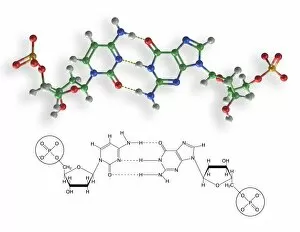
Background imageRibonucleic Acid Collection: Cytosine-guanine interaction, artwork C017 / 7215

Cytosine-guanine interaction, artwork C017 / 7215
Cytosine-guanine interaction. Computer artwork showing the structure of bound cytosine (left) and guanine molecules (right)

Background imageRibonucleic Acid Collection: Thymine molecule, artwork C017 / 7366

Thymine molecule, artwork C017 / 7366
Thymine molecule. Computer artwork showing the structure of a molecule of the nucleobase thymine. Atoms are colour-coded spheres: carbon (green), nitrogen (blue), oxygen (red), and hydrogen (white)

Background imageRibonucleic Acid Collection: Thymine molecule, artwork C017 / 7365

Thymine molecule, artwork C017 / 7365
Thymine molecule. Computer artwork showing the structure of a molecule of the nucleobase thymine. Atoms are colour-coded spheres: carbon (green), nitrogen (blue), oxygen (red), and hydrogen (white)

Background imageRibonucleic Acid Collection: Cytosine-guanine interaction, artwork C017 / 7216

Cytosine-guanine interaction, artwork C017 / 7216
Cytosine-guanine interaction. Computer artwork showing the structure of bound cytosine (left) and guanine molecules (right)

Background imageRibonucleic Acid Collection: Thymine-adenine interaction, artwork C017 / 7367

Thymine-adenine interaction, artwork C017 / 7367
Thymine-adenine interaction. Computer artwork showing the structure of bound thymine and adenine molecules. Atoms are shown as colour-coded spheres: carbon (green), hydrogen (white)

Background imageRibonucleic Acid Collection: Retrovirus, artwork F007 / 6437

Retrovirus, artwork F007 / 6437
Retrovirus, computer artwork. Retroviruses are viruses that have an RNA (ribonucleic acid) genome. They use reverse transcriptase to create a DNA (deoxyribonucleic acid)

Background imageRibonucleic Acid Collection: Glycine riboswitch molecule F007 / 9921

Glycine riboswitch molecule F007 / 9921
Molecular model of the bacterial glycine riboswitch. This is an RNA element that can bind the amino acid glycine. Glycine riboswitches usually consist of two metabolite-binding aptamer domains tandem

Background imageRibonucleic Acid Collection: Glycine riboswitch molecule F007 / 9906

Glycine riboswitch molecule F007 / 9906
Molecular model of the bacterial glycine riboswitch. This is an RNA element that can bind the amino acid glycine. Glycine riboswitches usually consist of two metabolite-binding aptamer domains tandem

Background imageRibonucleic Acid Collection: Human 80S ribosome F007 / 9902

Human 80S ribosome F007 / 9902
Ribosomal subunit. Computer model showing the structure of the RNA (ribonucleic acid) molecules in an 80S (large) ribosomal sub-unit. Ribosomes are composed of protein and RNA

Background imageRibonucleic Acid Collection: Human 80S ribosome F007 / 9898

Human 80S ribosome F007 / 9898
Ribosomal subunit. Computer model showing the structure of the RNA (ribonucleic acid) molecules in an 80S (large) ribosomal sub-unit. Ribosomes are composed of protein and RNA


All Professionally Made to Order for Quick Shipping

"Unraveling the Secrets of Ribonucleic Acid: The Double-Stranded RNA Molecule" In the intricate world of molecular biology, ribonucleic acid (RNA) takes center stage as a vital player in various biological processes. This captivating molecule, often overshadowed by its famous cousin DNA, holds immense potential and complexity. DNA transcription sets the stage for RNA's crucial role. As a double-stranded RNA molecule unwinds, it serves as a template to synthesize single-stranded messenger RNA (mRNA), carrying genetic information from the nucleus to the cytoplasm. A mesmerizing molecular model showcases this elegant dance of transcription. Within bacterial ribosomes, another fascinating aspect unfolds. These cellular factories decode mRNA sequences into proteins through translation—a fundamental process that sustains life itself. Peering into their microscopic world reveals an awe-inspiring view of these tiny machines at work. But not all encounters with RNA are beneficial; some bring about disease-causing agents like human respiratory syncytial virus or paramyxovirus particles. Through electron microscopy, we witness their hauntingly beautiful structures—reminders of nature's delicate balance between beauty and danger. Electrophoresis techniques allow scientists to analyze and separate different types of RNAs based on size and charge—an invaluable tool in unraveling their mysteries. Such experiments reveal intriguing patterns under UV light that hint at hidden secrets within these molecules' structure and function. The realm of RNA extends beyond mere replication; it undergoes editing too. Molecular models showcase specialized enzymes responsible for altering specific nucleotides within an RNA sequence—a testament to nature's ingenuity in fine-tuning genetic information. Ribonucleases further highlight the multifaceted nature of RNAs—their ability to degrade both RNA-DNA hybrids and pure forms with precision is truly remarkable. Visualizing this interaction provides insights into how cells regulate gene expression through controlled degradation mechanisms.