Home > Popular Themes > Human Body
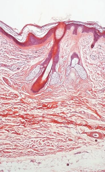
Human skin section, light micrograph P710 / 0472
![]()

Wall Art and Photo Gifts from Science Photo Library
Human skin section, light micrograph P710 / 0472
Human skin. Light micrograph of a section through healthy human skin. The outer surface of the skin is at top. The uppermost surface is the epidermis, which is topped by the dead stratum corneum layer. Cells are continually shed from this layer and are replaced by cells from the living underlying layer, the stratum germinativum (purple). This layer lies on top of the dermis, the thickest skin layer, which contains hair follicles (upper centre), sebaceous glands (one seen either side of the hair shaft), sweat glands (upper right) and blood vessels (for instance the red oval at lower right). The body of the dermis comprises fatty and connective tissue. Magnification: x59 when printed 10cm high
Science Photo Library features Science and Medical images including photos and illustrations
Media ID 9303769
© POWER AND SYRED/SCIENCE PHOTO LIBRARY
Dermis Epidermis Fatty Follicle Glands Hair Histological Histology Layers Sebaceous Gland Shaft Skin Stratum Corneum Surface Sweat Tissue Tissues Light Micrograph Light Microscope Stratum Germinativum
EDITORS COMMENTS
This print showcases a detailed light micrograph of a section through healthy human skin. The image reveals the intricate layers and structures that make up our largest organ. At the top, we see the outer surface of the skin, with the epidermis taking center stage. Topped by the dead stratum corneum layer, this constantly shedding layer is continuously replenished by cells from the living underlying layer known as the stratum germinativum. Moving deeper into the skin, we encounter various essential components such as hair follicles located in the upper center and sebaceous glands on either side of a hair shaft. Sweat glands can be observed towards the upper right portion of this fascinating composition. Additionally, blood vessels are visible throughout, including a distinct red oval at lower right. The dermis steals attention as it emerges as one of thickest layers within our skin structure. Comprising fatty and connective tissue, it provides support and nourishment to all other layers above it. With its vibrant colors and remarkable magnification capabilities (x59 when printed 10cm high), this print offers an awe-inspiring glimpse into both biology and anatomy. It serves as a testament to our complex biological makeup while highlighting key features like histology, sweat production, sebaceous gland function, and more – all contributing to maintaining normal healthy human skin.
MADE IN THE UK
Safe Shipping with 30 Day Money Back Guarantee
FREE PERSONALISATION*
We are proud to offer a range of customisation features including Personalised Captions, Color Filters and Picture Zoom Tools
SECURE PAYMENTS
We happily accept a wide range of payment options so you can pay for the things you need in the way that is most convenient for you
* Options may vary by product and licensing agreement. Zoomed Pictures can be adjusted in the Basket.

