Mounted Print > Popular Themes > Human Body
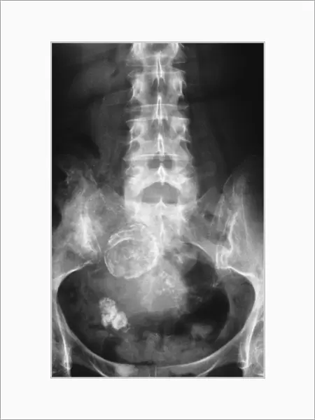
Mounted Print : Calcified cysts of the ovary
![]()

Mounted Prints from Science Photo Library
Calcified cysts of the ovary
Pelvic X-ray (front view) showing calcified cysts in a womans ovary (white, lower left)
Science Photo Library features Science and Medical images including photos and illustrations
Media ID 9242547
© GJLP - CNRI/SCIENCE PHOTO LIBRARY
Adult Patient Calcified Cyst Cysts Diagnosis Gynaecology Ovary Condition Disorder
10"x8" Mount with 8"x6" Print
Discover the intricacies of human anatomy with Media Storehouse's Mounted Photos featuring the Calcified Cysts of the Ovary. This captivating image, sourced from Science Photo Library, showcases a pelvic X-ray revealing the presence of calcified cysts in a woman's ovary. These mounted photos offer a unique and educational perspective, providing an up-close look into the medical world. Ideal for medical professionals, students, or anyone with an interest in anatomy, these high-quality mounted photos make for a thought-provoking addition to any office or learning space.
Printed on 8"x6" paper and suitable for use in a 10"x8" frame (frame not included). Prints are mounted with card both front and back. Featuring a custom cut aperture to match chosen image. Professional 234gsm Fujifilm Crystal Archive DP II paper.
Photo prints supplied in custom cut card mount ready for framing
Estimated Image Size (if not cropped) is 12.9cm x 20.3cm (5.1" x 8")
Estimated Product Size is 20.3cm x 25.4cm (8" x 10")
These are individually made so all sizes are approximate
Artwork printed orientated as per the preview above, with portrait (vertical) orientation to match the source image.
EDITORS COMMENTS
This print from Science Photo Library offers a unique glimpse into the intricate complexities of the female reproductive system. The image showcases a front view X-ray of a woman's pelvis, specifically highlighting calcified cysts within her ovary. These cysts, which appear as white masses in the lower left region, provide valuable insights into various gynecological conditions and disorders. The diagnosis of calcified cysts in the ovary is not uncommon among adult patients and can be associated with conditions such as polycystic ovarian syndrome (PCOS). This medical anomaly occurs when multiple small fluid-filled sacs develop within the ovaries, leading to hormonal imbalances and potential fertility issues. The photograph serves as an essential tool for healthcare professionals specializing in gynecology. It allows them to visually analyze and diagnose such conditions accurately, aiding in effective treatment plans for affected women. Moreover, it highlights the significance of ongoing research and advancements in understanding these complex disorders that impact countless individuals worldwide. Science Photo Library continues its commitment to providing high-quality visual resources that bridge the gap between scientific knowledge and public awareness. Through this remarkable image capturing calcified cysts of the ovary, they contribute to our collective understanding of human anatomy while emphasizing compassion-driven healthcare practices.
MADE IN THE UK
Safe Shipping with 30 Day Money Back Guarantee
FREE PERSONALISATION*
We are proud to offer a range of customisation features including Personalised Captions, Color Filters and Picture Zoom Tools
SECURE PAYMENTS
We happily accept a wide range of payment options so you can pay for the things you need in the way that is most convenient for you
* Options may vary by product and licensing agreement. Zoomed Pictures can be adjusted in the Basket.

