Jigsaw Puzzle : LM of nerve cells and fibres in brain tissue


![]()
Jigsaw Puzzles from Science Photo Library
LM of nerve cells and fibres in brain tissue
Brain tissue. Light micrograph of a section through the grey matter of a normal brain. There are several nerve cell bodies (objects in white areas) surrounded by associated dendrites and supportive glial cells. Some nuclei are visible as darker patches at the centre of the cell bodies. Dendrites, the smaller of the two types of nerve process, act as sensory receptors for the nerve cells. Grey matter in the brain consists of cell bodies, branched dendrites, glial cells and blood vessels. Magnification: x200 at 5x7cm size
Science Photo Library features Science and Medical images including photos and illustrations
Media ID 6448947
© PASIEKA/SCIENCE PHOTO LIBRARY
Cell Body Grey Matter Nerve Cell Nervous Neurone Neurone Cells System Brain Cells Light Micrograph
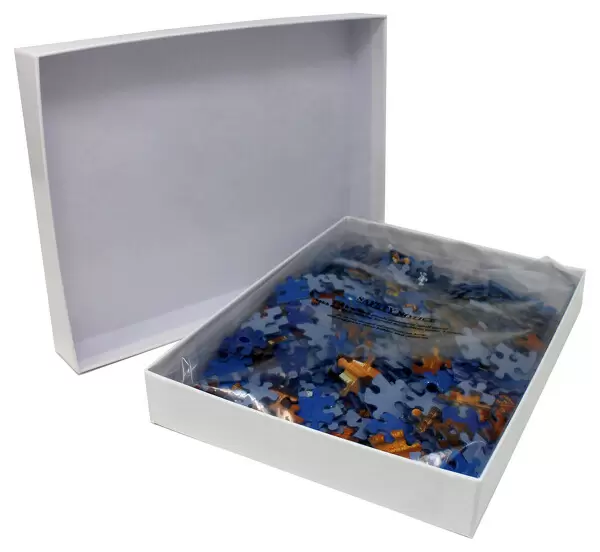
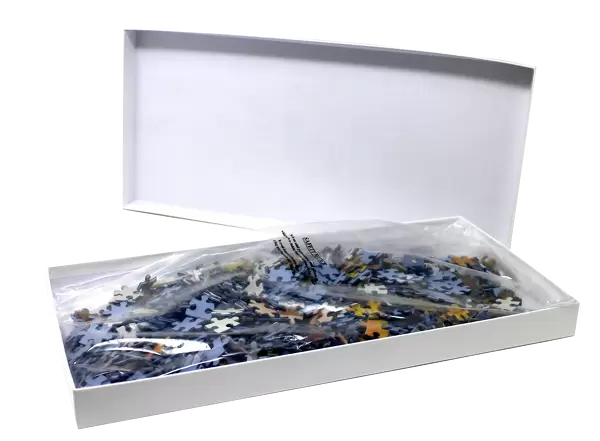
Jigsaw Puzzle (400 Pieces)
Discover the intricacies of the human brain with our captivating jigsaw puzzles from Media Storehouse. This scientific puzzle features a mesmerizing image of nerve cells and fibers in brain tissue, captured by the Science Photo Library. Assemble the pieces to reveal the complex network that forms the foundation of our thoughts, memories, and emotions. Engage your mind and ignite your curiosity with this challenging yet rewarding puzzle.
400 piece puzzles are custom made in the UK and hand-finished on 100% recycled 1.5 mm millboard. There is a level of repetition in jigsaw shapes with each matching piece away from its pair. The completed puzzle measures 31x47cm and is delivered packaged in an attractive presentation box specially designed to fit most letter box slots
Jigsaw Puzzles are an ideal gift for any occasion
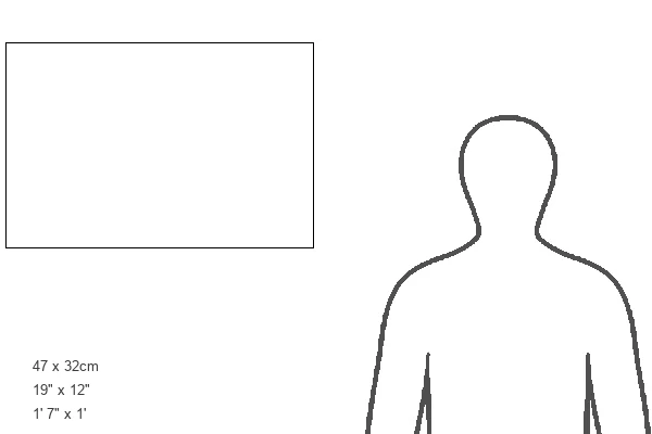
Estimated Product Size is 47.2cm x 31.5cm (18.6" x 12.4")
These are individually made so all sizes are approximate
Artwork printed orientated as per the preview above, with landscape (horizontal) or portrait (vertical) orientation to match the source image.
EDITORS COMMENTS
This print showcases the intricate beauty of nerve cells and fibers within brain tissue. Taken under a light microscope, the image reveals a section through the grey matter of a healthy brain. The white areas represent several nerve cell bodies, each surrounded by their associated dendrites and supportive glial cells. The darker patches at the center of these cell bodies signify visible nuclei, adding depth to this mesmerizing composition. Dendrites, which are sensory receptors for nerve cells, can be observed as smaller nerve processes branching out from the cell bodies. Grey matter in the brain consists not only of these remarkable cell bodies and branched dendrites but also includes glial cells and blood vessels that play crucial roles in maintaining proper neural function. With a magnification level of x200 at 5x7cm size, this photograph allows us to appreciate the complexity and interconnectedness present within our nervous system. It serves as a reminder of how intricately designed our brains are and highlights its importance in regulating various bodily functions. This stunning visual representation is brought to you by Science Photo Library – an invaluable resource for those seeking knowledge about human anatomy, specifically pertaining to neurology.
MADE IN THE UK
Safe Shipping with 30 Day Money Back Guarantee
FREE PERSONALISATION*
We are proud to offer a range of customisation features including Personalised Captions, Color Filters and Picture Zoom Tools
SECURE PAYMENTS
We happily accept a wide range of payment options so you can pay for the things you need in the way that is most convenient for you
* Options may vary by product and licensing agreement. Zoomed Pictures can be adjusted in the Basket.