Jigsaw Puzzle : False-colour SEM of a lobule of the liver


![]()
Jigsaw Puzzles from Science Photo Library
False-colour SEM of a lobule of the liver
False-colour scanning electron micrograph (SEM) of a lobule of the liver. Parenchyma tissue in the liver is arranged into lobules of cells, one of which is seen here. Each lobule has a central vein (black circle here); portal tracts at the boundary of each lobule (beyond this field of view) lead blood to flow along paths or sinusoids (grey) between hepatic cells, towards the centre of the lobule. In the process, blood is detoxified of waste products; proteins are metabolized; blood clotting agents, vitamin A and bile are synthe- sized; and spent red blood cells are reabsorbed. Magnification: x570 at 6x7cm size. Magnification: x875 at 4x5 inch size
Science Photo Library features Science and Medical images including photos and illustrations
Media ID 6449669
© PROF. P. MOTTA/DEPT. OF ANATOMY/UNIVERSITY LA SAPIENZA , ROME/SCIENCE PHOTO LIBRARY
Hepatic Liver Lobule Lobules False Coloured
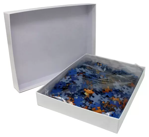
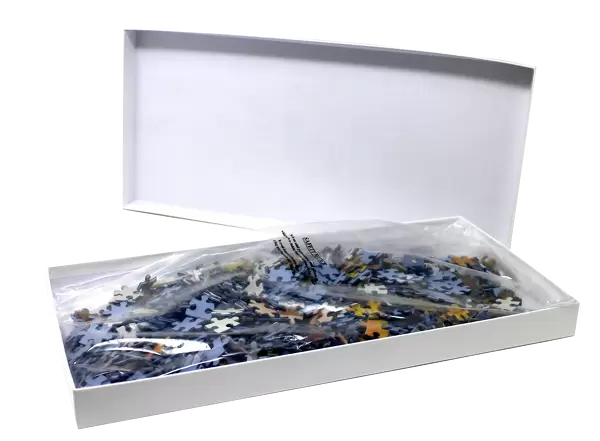
Jigsaw Puzzle (500 Pieces)
Discover the wonders of the human body with Media Storehouse's Jigsaw Puzzles. Our latest addition to the collection is a captivating False-colour SEM of a lobule of the liver, courtesy of Science Photo Library. Explore the intricate details of this vital organ's parenchyma tissue, arranged into lobules of cells, and piece together the mystery of nature. Engage in a challenging yet rewarding puzzle experience that brings science to life right at your fingertips.
500 piece puzzles are custom made in the UK and hand-finished on 100% recycled 1.5 mm millboard. There is a level of repetition in jigsaw shapes with each matching piece away from its pair. The completed puzzle measures 38x50cm and is delivered packaged in an attractive presentation box specially designed to fit most letter box slots
Jigsaw Puzzles are an ideal gift for any occasion
Estimated Product Size is 50.2cm x 38cm (19.8" x 15")
These are individually made so all sizes are approximate
Artwork printed orientated as per the preview above, with landscape (horizontal) or portrait (vertical) orientation to match the source image.
EDITORS COMMENTS
This print showcases a false-colour scanning electron micrograph (SEM) of a lobule in the liver. The intricate parenchyma tissue is organized into lobules, and this particular image offers an up-close view of one such structure. At the center of the lobule, we can observe a prominent black circle representing the central vein. Beyond our field of view lie portal tracts that demarcate each lobule's boundary. These portal tracts play a crucial role in directing blood flow along sinusoids or paths between hepatic cells towards the center of the lobule, where detoxification and various metabolic processes occur. Within these sinuous pathways, blood undergoes essential transformations: waste products are eliminated, proteins are metabolized, clotting agents are synthesized alongside vitamin A and bile production. Additionally, spent red blood cells find their final destination as they are reabsorbed within this intricate network. The level of detail captured by this SEM is truly remarkable; it magnifies at x570 for 6x7cm size prints and x875 for 4x5 inch size prints. This stunning visual representation provides valuable insights into liver anatomy while highlighting its vital functions within the human body.
MADE IN THE UK
Safe Shipping with 30 Day Money Back Guarantee
FREE PERSONALISATION*
We are proud to offer a range of customisation features including Personalised Captions, Color Filters and Picture Zoom Tools
SECURE PAYMENTS
We happily accept a wide range of payment options so you can pay for the things you need in the way that is most convenient for you
* Options may vary by product and licensing agreement. Zoomed Pictures can be adjusted in the Basket.