
Lobule Collection
A lobule, a fascinating structure found in various parts of the human body, plays a crucial role in their respective functions


All Professionally Made to Order for Quick Shipping
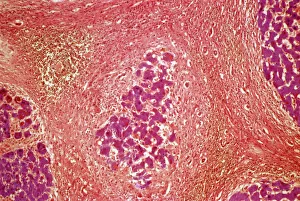
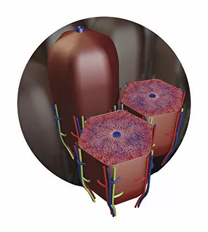
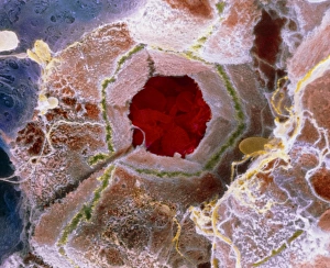
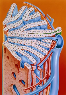
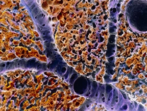
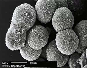
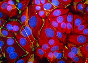
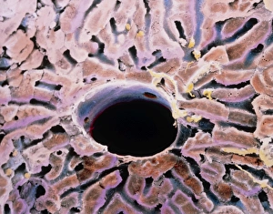
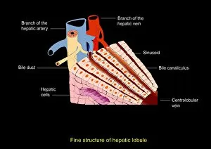
A lobule, a fascinating structure found in various parts of the human body, plays a crucial role in their respective functions. In the cross-section diagram of the human ear, we can observe tiny lobules responsible for transmitting sound signals to our brain. Moving on to liver tissue cirrhosis, a light micrograph reveals damaged lobules due to chronic liver disease. Shifting our focus to the thyroid gland capillaries and blood vessels captured through scanning electron microscopy (SEM), we witness intricate networks supporting this vital endocrine organ's function. These microscopic structures within the thyroid gland are known as lobules. As we explore further into breast anatomy, an intriguing cross-section image showcases numerous components such as ribs, pectoral muscles, lungs, fatty tissues, blood vessels, and most importantly - lobules. These milk-producing units play a significant role during lactation and breastfeeding. The biomedical illustration of liver lobule provides us with valuable insights into its complex architecture. This structural arrangement allows efficient processing of nutrients and detoxification within this essential organ. Intriguingly enough, even French line engravings from the 19th century depict these remarkable formations found throughout our bodies. The detailed illustrations highlight how early anatomists recognized and appreciated the significance of these small but mighty structures called lobules. Understanding different aspects of human physiology is incomplete without acknowledging these specialized units present in organs like ears or breasts. Whether it be hearing or nourishing newborns with breast milk – all made possible by these incredible little entities known as lobules.