Fraud Blocker Skip to main content

Bones Collection

"Bones: Unveiling the Hidden Structures of Life" Delving into the depths of human anatomy

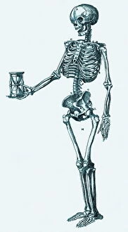
Background imageBones Collection: Human skeleton

Human skeleton. Historical artwork of a human skeleton holding an hourglass. The 206 bones of the skeleton provide protection and support to the body

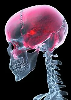
Background imageBones Collection: Headache, X-ray artwork

Headache, X-ray artwork
Headache. Computer X-ray artwork of a lateral view of a human skull. The red area represents pain caused by a headache (cephalalgia)

Background imageBones Collection: Skeleton of a greyhound

Skeleton of a greyhound
Detailed anatomical drawing by Raymond Sheppard in pen and ink of the skeleton of a greyhound, with bones fully labelled

Background imageBones Collection: The Young Ones during filming in Bristol. Starring Christopher Ryan as Mike

The Young Ones during filming in Bristol. Starring Christopher Ryan as Mike, Rik Mayall as Rick, Nigel Planer as Neil and Adrian Edmondson as Vyvyan. August 1982

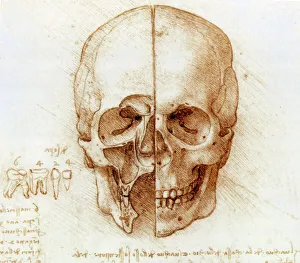
Background imageBones Collection: Skull anatomy by Leonardo da Vinci

Skull anatomy by Leonardo da Vinci. Historical artwork and notes on the anatomy of the human skull and teeth, by the Italian artist and scientist Leonardo da Vinci (1452-1519)

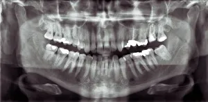
Background imageBones Collection: Panoramic dental X-ray

Panoramic dental X-ray showing all the teeth in the upper and lower jaws

Background imageBones Collection: Broken wrist bone, X-ray C017 / 7187

Broken wrist bone, X-ray C017 / 7187
Broken wrist bone. X-ray of the hand of a 12 year old male with a fractured trapezium bone. The trapezium is the leftmost bone in the group of bones at the base of the bad

Background imageBones Collection: Knapped Flint Tools

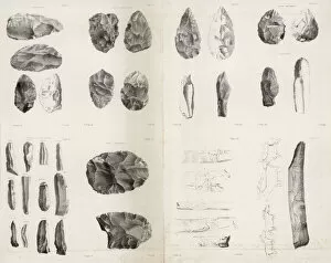
Knapped Flint Tools
A selection of flint and bones tools from a range of prehistoric eras incl. microliths used in compound tools of the mesolithic to earlier scrapers and choppers

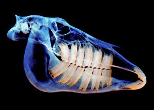
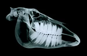
Background imageBones Collection: Horses skull

Horses skull, coloured X-ray

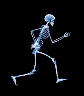
Background imageBones Collection: Skeleton playing rugby

Skeleton playing rugby. Computer enhanced X- ray of a skeleton running with a rugby ball

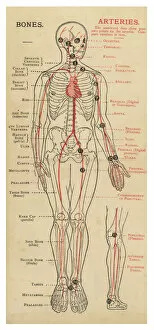
Background imageBones Collection: Human body with bones and arteries

Human body with bones and arteries
A diagram of the human body, with details of bones and arteries

Background imageBones Collection: Chef Andrew Schillar gives a dog a bone from the side entrance of his kitchen. undated

Chef Andrew Schillar gives a dog a bone from the side entrance of his kitchen. undated

Background imageBones Collection: Skeletons, X-ray artwork

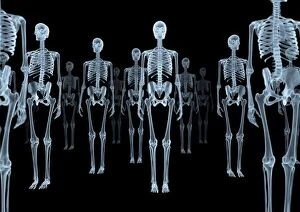
Skeletons, X-ray artwork
Skeleton crowd. Computer X-ray artwork of a group of human skeletons viewed from the front

Background imageBones Collection: Full body scan, MRI scan

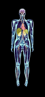
Full body scan, MRI scan
Full body scan of a female swimmer. Her brain is purple (top), her heart and arteries are red/orange (upper centre), her kidneys are pale green (centre)

Background imageBones Collection: Death as Assassin, 1851 (wood engraving)

Death as Assassin, 1851 (wood engraving)
XJF143235 Death as Assassin, 1851 (wood engraving) by Rethel, Alfred (1816-59); Private Collection; (add.info.: the outbreak of cholera at the Masked Ball in Paris; ); German, out of copyright

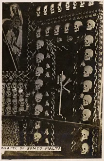
Background imageBones Collection: Ossuary Chapel of skulls - Valletta, Malta

Ossuary Chapel of skulls - Valletta, Malta - destroyed by a bomb during World War II. Date: circa 1910s

Background imageBones Collection: Skeleton drinking, X-ray

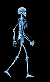
Skeleton drinking, X-ray
Human skeleton walking. Computer artwork based on an X-ray of a full human skeleton walking. The 206 bones provide protection & structural support, and the joints allow locomotion

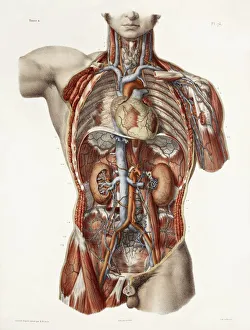
Background imageBones Collection: Cardiovascular system, historical artwork

Cardiovascular system, historical artwork. 19th Century hand coloured lithographic print showing the blood vessels (red and blue) of the human neck and torso

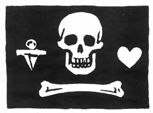
Background imageBones Collection: PIRATES: JOLLY ROGER FLAG. Flag of the English pirate, Stede Bonnet

PIRATES: JOLLY ROGER FLAG. Flag of the English pirate, Stede Bonnet

Background imageBones Collection: Cat Skeleton

Cat Skeleton
LA-82 Cat Skeleton Jean Michel Labat contact details: prints@ardea.com tel: +44 (0) 20 8318 1401

Background imageBones Collection: Dog skull X-ray

Dog skull X-ray

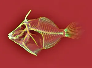
Background imageBones Collection: Triggerfish skeleton, X-ray

Triggerfish skeleton, X-ray
Triggerfish skeleton. Coloured X-ray of a triggerfish (family Balistidae). Its name originates from its ability to erect the first two dorsal spines (upper left), which can then lock or unlock

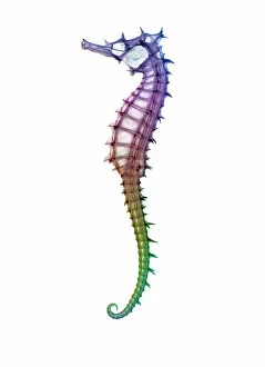
Background imageBones Collection: Seahorse skeleton

Seahorse skeleton. Coloured x-ray of a seahorse (Hippocampus sp.). Seahorse populations are under threat due to the fishing industry

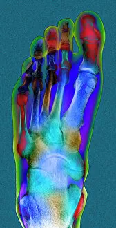
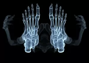
Background imageBones Collection: Normal foot, X-ray

Normal foot, X-ray
Normal foot. Coloured X-ray of a healthy human foot seen from the top (antero-posterior or dorso- plantar projection). The foot comprises 26 bones and two sesamoid bones (not seen)

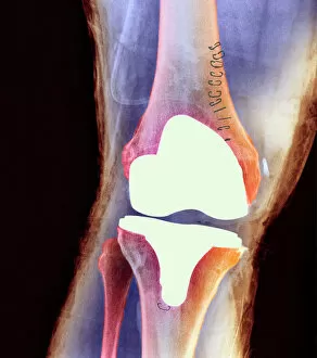
Background imageBones Collection: Knee joint prosthesis, X-ray

Knee joint prosthesis, X-ray
Knee joint replacement. Coloured X-ray of the knee of a 56 year old man (front view) showing an artificial (prosthetic) joint replacing the knee joint

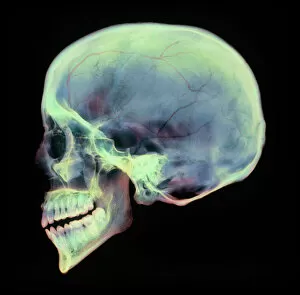
Background imageBones Collection: Human skull, X-ray

Human skull, X-ray
Human skull. Coloured X-ray of a sagittal section through a human skull. The skull has been sliced in half down the centre, revealing details of the internal structures

Background imageBones Collection: Horse skull

Horse skull (Equus caballus), side view X-ray

Background imageBones Collection: Bunions, X-ray

Bunions, X-ray
Bunions. Coloured X-ray of an elderly womans deformed inward-turning toes due to bunions (orange, lower centre). This deformity (hallux valgus) is caused by wearing ill-fitting shoes

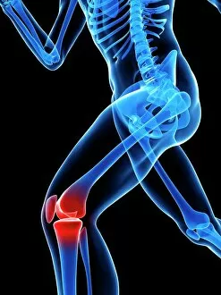
Background imageBones Collection: Knee pain, conceptual artwork

Knee pain, conceptual artwork

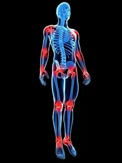
Background imageBones Collection: Joint pain, conceptual artwork

Joint pain, conceptual artwork

Background imageBones Collection: Total hip replacement, X-ray

Total hip replacement, X-ray
Total hip replacement. Coloured frontal X-ray of a female pelvis with a total hip replacement (white, lower right). The replacement hip consists of a ball and shaft

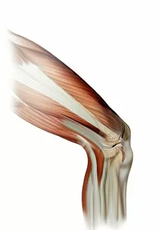
Background imageBones Collection: Damaged knee ligament, artwork

Damaged knee ligament, artwork
Damaged knee ligament. Computer artwork of the muscle structure of the leg and anatomy of the human knee joint, showing a tear (dark line, centre right) in one of the cruciate ligaments (beige)

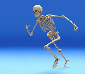
Background imageBones Collection: Running skeleton in body, artwork

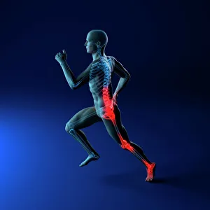
Running skeleton in body, artwork
Running skeleton inside an outline of the body, computer artwork

Background imageBones Collection: Skateboarder 1970S

Skateboarder 1970S
A boy skateboarding in protecive gear, including shin pads and a helmet, Crawley, Sussex, England

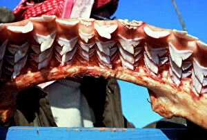
Background imageBones Collection: Jaw of a 4 metre tiger shark, replaceable teeth in 9 rows Egypt Red Sea

Jaw of a 4 metre tiger shark, replaceable teeth in 9 rows Egypt Red Sea
KA-516 Jaw of a 4 metre tiger shark, replaceable teeth in 9 rows Egypt Red Sea Kurt Amsler Please note that prints are for personal display purposes only and may not be reproduced in any way

Background imageBones Collection: Skeleton playing football

Skeleton playing football. Computer enhanced X- ray of a skeleton bouncing a football off his knee

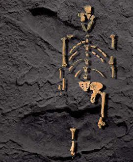
Background imageBones Collection: Footprints and skeleton of Lucy

Footprints and skeleton of Lucy (Australopithecus afarensis). The fossilised bones of the A. afarensis specimen known as Lucy are superimposed over footprints made by the same species. A

Background imageBones Collection: Skeleton & Ligaments

Skeleton & Ligaments
Physiological diagram of the skeleton and ligaments

Background imageBones Collection: Upper body skeleton, computer artwork

Upper body skeleton, computer artwork
Upper body skeleton. Computer artwork of a human skeleton showing the bones of the upper body. At the top is the base of the skull and the jaw bone

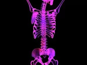
Background imageBones Collection: Skeleton from below, X-ray artwork

Skeleton from below, X-ray artwork
Skeleton. Computer x-ray artwork of a human skeleton viewed from below

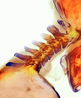
Background imageBones Collection: Neck vertebrae extended, X-ray

Neck vertebrae extended, X-ray
Bending of the neck. Coloured X-ray of a side view of the neck of a man showing extension of healthy cervical vertebrae (bones)

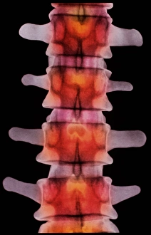
Background imageBones Collection: Coloured X-ray of lumbar vertebrae of the spine

Coloured X-ray of lumbar vertebrae of the spine
Lumbar spine. Coloured X-ray of four lumbar vertebrae of the human spine, seen in antero- posterior view. Five lumbar bones are found in the lower back

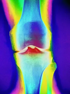
Background imageBones Collection: Coloured X-ray of a human knee joint

Coloured X-ray of a human knee joint
Knee joint. Coloured X-ray of a healthy human knee joint. Two bones meet at the knee forming a joint that works like a hinge

Background imageBones Collection: Skeleton on a penny farthing

Skeleton on a penny farthing. Composite x-ray of a skeleton riding a penny farthing bicycle represented by the coins it is named after

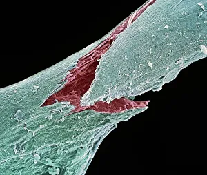
Background imageBones Collection: Osteoporotic bone, SEM

Osteoporotic bone, SEM
Osteoporotic bone. Coloured scanning electron micrograph (SEM) of fractured bone tissue from a patient suffering from osteoporosis (brittle bone disease)

Background imageBones Collection: Running injuries, conceptual artwork

Running injuries, conceptual artwork

Background imageBones Collection: Fractured ankle, X-ray

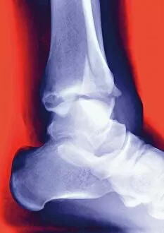
Fractured ankle, X-ray
Fractured ankle. Coloured profile X-ray of a distal fibula fracture (upper centre). The fibula is the smaller leg bone running down from top centre. The larger bone is the tibia

Background imageBones Collection: Sahelanthropus tchadensis skull

Sahelanthropus tchadensis skull. Artwork of a reconstruction of the Toumai skull, one of only a small number of fossils of the hominin Sahelanthropus tchadensis


All Professionally Made to Order for Quick Shipping

"Bones: Unveiling the Hidden Structures of Life" Delving into the depths of human anatomy, Leonardo da Vinci's "Skull Anatomy" showcases the intricate beauty and complexity of bones. A graceful greyhound's skeleton reveals the elegance and agility that lies beneath its sleek exterior. Behind-the-scenes in Bristol, Christopher Ryan transforms into Mike from "The Young Ones, " a character whose humor is as sharp as his bone structure. Captured on an X-ray, a broken wrist bone serves as a reminder of our vulnerability and resilience in the face of injury. The majestic skull of a horse stands as a testament to their strength and power, reminding us of their historical significance alongside humans. Even skeletons enjoy some rough play. Witnessing a rugby-playing skeleton brings forth both amusement and admiration for these resilient structures. Knapped Flint Tools take us back in time, showcasing how early humans utilized bones to shape tools for survival and progress. Peering through panoramic dental X-rays unravels secrets hidden within our mouths – teeth anchored by strong bones supporting our smiles. Our bodies are marvels intertwined with arteries that nourish every cell while sturdy bones provide support - truly works of art designed by nature itself. In an act of kindness, Chef Andrew Schillar shares his culinary delight with man's best friend - offering them not just food but also joyous moments gnawing on a bone outside his kitchen sanctuary. Illustrated through wood engraving in 1851, "Death as Assassin" reminds us that even though life may be fleeting, it is ultimately connected to the skeletal framework we all share. Modern technology unveils our innermost secrets with full-body scans like MRI scans – exposing not only flesh but also revealing the silent symphony orchestrated by our interconnected bones.