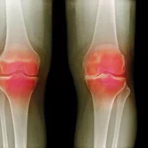
Knee joint prosthesis, X-ray

![]()
Wall Art and Photo Gifts from Science Photo Library
Knee joint prosthesis, X-ray
Knee joint replacement. Coloured X-ray of the knee of a 56 year old man (front view) showing an artificial (prosthetic) joint replacing the knee joint. The entire knee joint surface has been replaced here. The implant (white) made of metal alloy attaches to the bottom of the femur (thigh bone, upper frame) and to the top of the tibia (shin bone, lower frame). It forms a flexible joint that can hinge like the old joint, relieving joint pain and immobility. Following surgery, metal sutures (blue) are seen on the skin surface and a catheter draining the area is visible. Knee joint replacement is conducted usually because of joint disease such as osteoarthritis or due to injury
Science Photo Library features Science and Medical images including photos and illustrations
Media ID 6431755
© SCIENCE PHOTO LIBRARY
Arthritic Arthritis Bones Femur Fibula Front Frontal View Implant Implants Metal Alloy Musculoskeletal Disorders Osteo Arthritis Patient Prosthesis Prosthetic Device Radiography Replaced Replacement Shin Sutures Thigh Tibia Total Treatment Wound False Coloured Health Care Staples
EDITORS COMMENTS
This print showcases a knee joint prosthesis, providing an intricate view of a knee joint replacement. The image captures the front view of a 56-year-old man's knee, where an artificial (prosthetic) joint has replaced the entire surface of the original knee joint. The implant, made from a durable metal alloy, securely attaches to both the femur (thigh bone) and tibia (shin bone), forming a flexible hinge-like joint that mimics natural movement. The purpose behind this innovative procedure is to alleviate excruciating joint pain and restore mobility for individuals suffering from conditions like osteoarthritis or severe injuries. Following surgery, visible metal sutures in blue can be observed on the skin surface while a catheter drains excess fluid from the area. This remarkable medical intervention not only highlights advancements in modern medicine but also emphasizes its impact on improving patients' quality of life. By replacing damaged joints with prosthetic devices, individuals can regain their ability to move freely without experiencing debilitating pain. Science Photo Library presents this false-colored X-ray image as part of their extensive collection dedicated to showcasing various aspects of health care and musculoskeletal disorders. It serves as a powerful visual representation of how medical science continues to evolve and provide effective solutions for those facing physical challenges related to their joints.
MADE IN THE UK
Safe Shipping with 30 Day Money Back Guarantee
FREE PERSONALISATION*
We are proud to offer a range of customisation features including Personalised Captions, Color Filters and Picture Zoom Tools
SECURE PAYMENTS
We happily accept a wide range of payment options so you can pay for the things you need in the way that is most convenient for you
* Options may vary by product and licensing agreement. Zoomed Pictures can be adjusted in the Basket.