Glass Place Mat : Dislocated elbow joint, X-ray
![]()

Home Decor from Science Photo Library
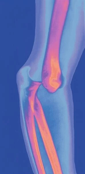
Dislocated elbow joint, X-ray
Dislocated elbow. Coloured X-ray of the bones (pink/orange) in a dislocated elbow joint. The humerus (upper arm bone, upper frame) has moved out of its correct position. It is normally in contact with the ends of the lower arm bones, the ulna (lower frame, left) and the radius. A dislocation is usually accompanied by tearing of the ligaments that hold the bones in place, which causes severe pain. A dislocation restricts or prevents the movement of the joint and causes swelling. If no fractures are present, the joint should be manipulated back into place and immobilized to allow the ligaments to heal
Science Photo Library features Science and Medical images including photos and illustrations
Media ID 6423783
© DR LINDA STANNARD, UCT/SCIENCE PHOTO LIBRARY
Bones Dislocated Dislocation Elbow Humerus Injured Injury Joint Radius Skeletal Ulna Condition Disorder Health Care
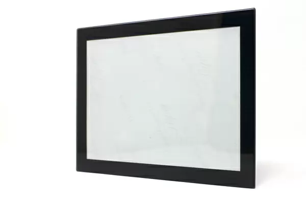
Glass Place Mat (Set of 4)
Set of 4 Glass Place Mats. Stylish and elegant polished safety glass, toughened and heat resistant (275x225mm, 7mm thick). Matching Coasters also available.
Set of 4 Glass Place Mats. Elegant polished safety glass and heat resistant. Matching Coasters may also be available
Estimated Image Size (if not cropped) is 12.5cm x 25.4cm (4.9" x 10")
Estimated Product Size is 22.5cm x 27.5cm (8.9" x 10.8")
These are individually made so all sizes are approximate
EDITORS COMMENTS
This print from Science Photo Library showcases a dislocated elbow joint in all its intricate detail. The vibrant pink and orange hues highlight the bones of the elbow, revealing the unsettling sight of the humerus, or upper arm bone, displaced from its correct position. Normally, this bone would be in contact with the ends of the ulna and radius – two lower arm bones that form part of this complex joint. A dislocation such as this is not only visually striking but also incredibly painful. It often involves tearing of ligaments responsible for holding these bones together. As a result, movement becomes restricted or even impossible while swelling ensues. In cases where no fractures are present, treatment typically involves manipulating the joint back into place followed by immobilization to allow for proper healing of ligaments. This image serves as a stark reminder of both our vulnerability to injury and medicine's ability to address such skeletal disorders. Science Photo Library has once again captured an extraordinary moment within medical science through their lens. This photograph stands as a testament to their dedication in providing invaluable visual resources for healthcare professionals and researchers alike.
MADE IN THE UK
Safe Shipping with 30 Day Money Back Guarantee
FREE PERSONALISATION*
We are proud to offer a range of customisation features including Personalised Captions, Color Filters and Picture Zoom Tools
SECURE PAYMENTS
We happily accept a wide range of payment options so you can pay for the things you need in the way that is most convenient for you
* Options may vary by product and licensing agreement. Zoomed Pictures can be adjusted in the Basket.


