
Ulna Collection
The ulna, one of the two bones in the forearm, plays a crucial role in our arm's structure and functionality


All Professionally Made to Order for Quick Shipping
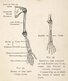
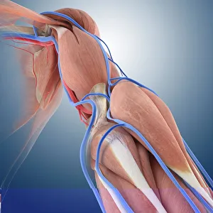
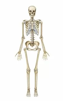
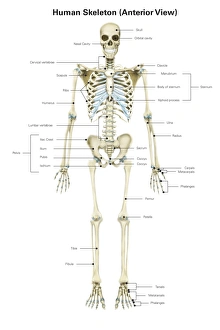
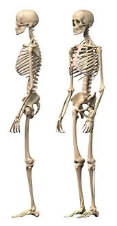
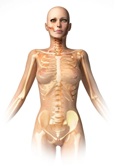
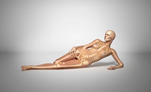
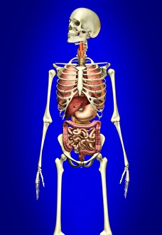
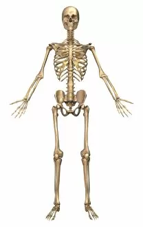
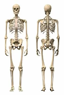
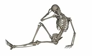
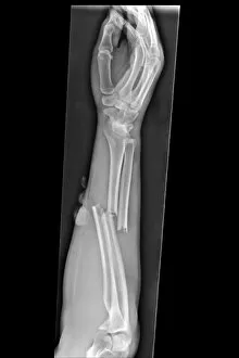
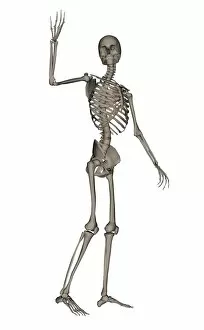
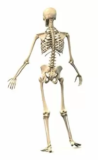
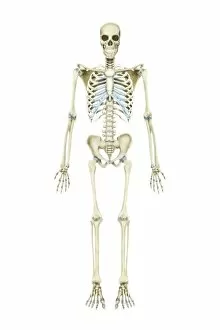
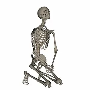
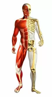
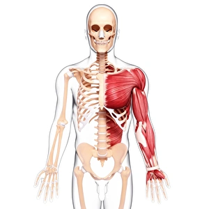
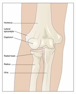
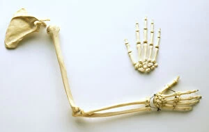
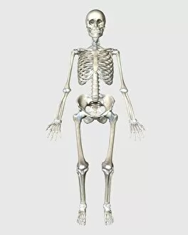
The ulna, one of the two bones in the forearm, plays a crucial role in our arm's structure and functionality. Its position alongside the radius allows for various movements, making it an essential component of our upper limb. With diagrams showcasing the intricate bones of the hand and arm, we can better understand how the ulna fits into this complex system. These visual aids provide a clear picture of its location and relationship with neighboring bones. Exploring arm circulation through anatomical artwork further emphasizes the significance of this bone. The ulna contributes to maintaining proper blood flow throughout our arms, ensuring optimal functioning. In contrast, images depicting a broken arm in a plaster cast or X-ray highlight situations where damage has occurred to this vital bone. Understanding these injuries helps medical professionals diagnose and treat fractures effectively. Examining different views of human skeletal systems provides valuable insights into how the ulna interacts with other bones within our bodies. From front view to side view and perspective view, these illustrations offer comprehensive knowledge about its placement and function. Not limited to humans alone, even animals like eagles possess their own version of an ulna bone. Engravings depicting an eagle's skeleton showcase similarities between species while highlighting unique adaptations specific to each creature. Furthermore, skiagrams capturing a child's hand reveal intricate details that might otherwise go unnoticed by naked eyes. These black-and-white photographs allow us to appreciate both fragility and resilience present within such small structures. By superimposing internal organs onto female body skeletons, we gain insight into how interconnected our bodies truly are. This integration highlights just how integral each individual bone is for overall health and well-being. Whether studying anatomy or biology or exploring topics related to fractures or close-ups on color backgrounds – all aspects contribute towards unraveling mysteries surrounding this remarkable bone: The Ulna. So next time you examine your own forearm or come across any reference related to arms' anatomy – take a moment to appreciate the ulna and its role in our everyday lives.