
Dislocated Collection
"Unveiling the Tales of Dislocation: A Journey through Medical History" Step into the realm of dislocation, where stories of resilience and innovation unfold


All Professionally Made to Order for Quick Shipping
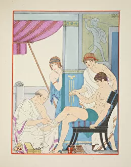
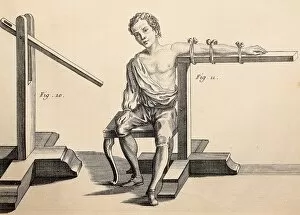
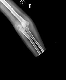
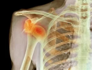
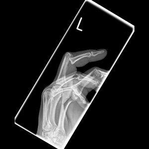
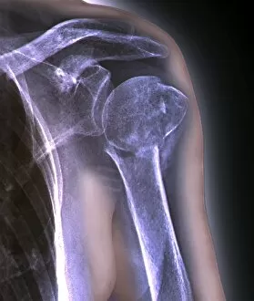
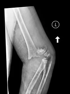
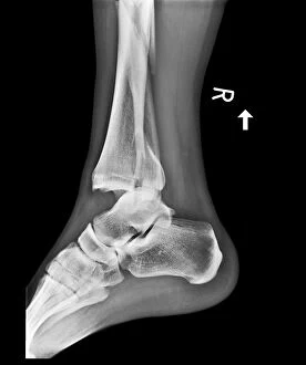
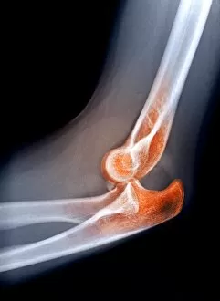
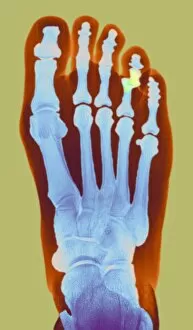
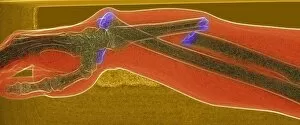
"Unveiling the Tales of Dislocation: A Journey through Medical History" Step into the realm of dislocation, where stories of resilience and innovation unfold. From wounded soldiers to ancient illustrations from The Works of Hippocrates, witness the evolution of medical practices. Explore a world filled with surgical equipment, including machines designed for precision and efficiency. Marvel at the 18th-century surgical knee pad and dislocation machine that revolutionized orthopedic care. Fractures and accidents captured in engravings remind us of the fragility of our bodies. Witness Luxation sous coracoidienne de l'épaule, Deplacement de la tête de l'humérus in vibrant color lithography, showcasing both artistry and anatomical accuracy. Delve deeper into history as you encounter equipment specifically crafted for dislocated arms. These images from ancient books transport us back to an era when healing was both science and art. Witness the power of pulleys as they aid in reducing dislocated thighs - a technique documented in The Household Physician published around 1898. This glimpse into past medical practices highlights humanity's relentless pursuit to restore mobility. Immerse yourself once again in a display featuring surgical tools alongside an illustration depicting bones within a human hand with a dislocated thumb. Here lies evidence that even centuries ago, physicians sought solutions for such common injuries. Finally, marvel at Roman ingenuity with their extension mechanism designed to treat dislocated knees. This testament to ancient engineering showcases how civilizations throughout time have grappled with similar challenges faced by modern medicine today. As we journey through these captivating glimpses into medical history, let us appreciate those who paved the way towards understanding and treating dislocations – forever changing lives one reduction at a time.