Glass Place Mat : Cats tooth, SEM


![]()
Home Decor from Science Photo Library
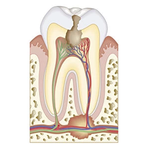
Cats tooth, SEM
Cats tooth. Coloured scanning electron micrograph (SEM) of a vertical fracture through a canine tooth of a domestic cat (Felis catus). At the centre of the tooth (dark coloured oval) is the pulp cavity, containing specialized connective tissue and sensory nerve fibres (dental pulp). The bulk of the tooth is dentine (green), a mineralised tissue with a similar composition to bone. Within the dentine are channels (dental tubules), containing odontoblasts, cells which produce new dentine (not shown). Surrounding the dentine is enamel (brown), the hardest substance in the body. If damage occurs to the enamel, self-repair is limited
Science Photo Library features Science and Medical images including photos and illustrations
Media ID 6451013
© STEVE GSCHMEISSNER/SCIENCE PHOTO LIBRARY
Brown Canine Dental Dentine Enamel False Colour Felis Catus Fracture Fractured Tooth False Coloured Section Sectioned
Glass Place Mat (Set of 4)
Set of 4 Glass Place Mats. Stylish and elegant polished safety glass, toughened and heat resistant (275x225mm, 7mm thick). Matching Coasters also available.
Set of 4 Glass Place Mats. Elegant polished safety glass and heat resistant. Matching Coasters may also be available
Estimated Image Size (if not cropped) is 18.4cm x 25.4cm (7.2" x 10")
Estimated Product Size is 22.5cm x 27.5cm (8.9" x 10.8")
These are individually made so all sizes are approximate
EDITORS COMMENTS
This print showcases the intricate beauty of a cat's tooth, captured through a coloured scanning electron microscope (SEM). The image reveals a vertical fracture that runs through the canine tooth of a domestic cat. At the heart of this dental marvel lies the pulp cavity, housing specialized connective tissue and sensory nerve fibers known as dental pulp. The bulk of the tooth is composed of dentine, which exhibits a striking green hue reminiscent of bone. Within this mineralized tissue are channels called dental tubules, containing odontoblasts – cells responsible for generating new dentine. Enveloping the dentine is enamel, boasting an alluring brown shade and recognized as one of nature's toughest substances. Delving into its biological significance, this photograph sheds light on the anatomy and structure underlying feline teeth. It serves as a testament to their remarkable adaptability in hunting and survival within their natural habitats. However, it also highlights that if damage occurs to the enamel layer, self-repair mechanisms are limited. Through this mesmerizing visual exploration at microscopic levels, we gain insight into both zoological wonders and dental biology alike. This extraordinary image from Science Photo Library invites us to appreciate nature's intricacies while deepening our understanding of these incredible creatures - Felis catus - who share our lives with such grace and mystery.
MADE IN THE UK
Safe Shipping with 30 Day Money Back Guarantee
FREE PERSONALISATION*
We are proud to offer a range of customisation features including Personalised Captions, Color Filters and Picture Zoom Tools
SECURE PAYMENTS
We happily accept a wide range of payment options so you can pay for the things you need in the way that is most convenient for you
* Options may vary by product and licensing agreement. Zoomed Pictures can be adjusted in the Basket.