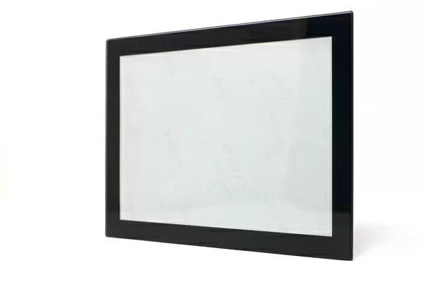
Glass Frame : Tea leaf, light micrograph


![]()
Mounted Prints from Science Photo Library
Tea leaf, light micrograph
Tea leaf. Light micrograph of a cross-section through a tea (Camellia sinensis) leaf. The upper and lower epidermis on the surfaces of the leaf are blue. Under the upper epidermis are palisade cells (brown), which contain chloroplasts, the site of photosynthesis. Beneath this a spongy mesophyll layer with large spaces between the cells. At bottom left, a stoma (pore) is seen. Stomata allow gases and water to enter and leave the plant. Magnification: x230 when printed 10 centimetres wide
Science Photo Library features Science and Medical images including photos and illustrations
Media ID 6300639
© DR KEITH WHEELER/SCIENCE PHOTO LIBRARY
Camellia Sinensis Chloroplasts Cross Section Epidermis Mesophyte Phloem Plant Anatomy Pore Pores Stoma Stomata Transverse Section Vascular Bundle Xylem Cells Light Micrograph Light Microscope Sectioned
8"x6" Glass Mount
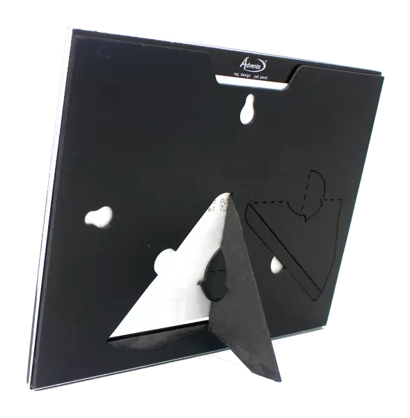
Wall mounted or free-standing, these black edged glass frames feature a smooth chamfered edge and a stylish black border (on back face of the glass). Manufactured from 4mm thick glass, Glass Mounts are a durable, professional way of displaying and protecting your prints. Your 8x6 print is slotted into the back of the frame so can easily be changed if needed.
Tempered Glass Mounts are ideal for wall display, plus the smaller sizes can also be used free-standing via an integral stand
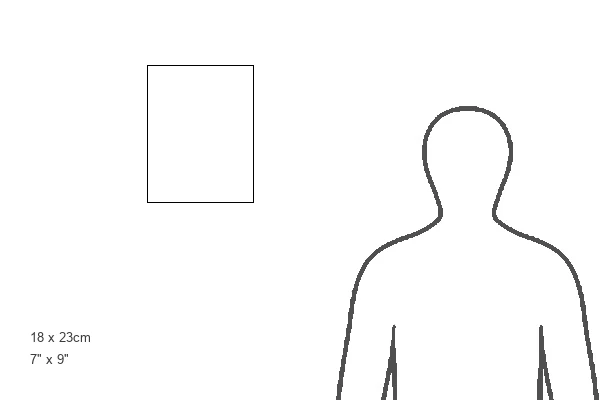
Estimated Image Size (if not cropped) is 15.2cm x 20.3cm (6" x 8")
Estimated Product Size is 17.7cm x 22.8cm (7" x 9")
These are individually made so all sizes are approximate
Artwork printed orientated as per the preview above, with portrait (vertical) orientation to match the source image.
EDITORS COMMENTS
This print showcases the intricate beauty of a tea leaf, captured under the lens of a light microscope. The cross-section of the leaf reveals its fascinating anatomy and highlights its vital role in photosynthesis. The upper and lower epidermis, depicted in shades of blue, form protective layers on either side of the leaf. Beneath the upper epidermis lies a layer of palisade cells, distinguished by their rich brown coloration. These cells house chloroplasts, which are responsible for capturing sunlight and converting it into energy through photosynthesis. Just below this layer is the spongy mesophyll, characterized by large spaces between its cells. Intriguingly positioned at the bottom left corner is a stoma or pore. Stomata play a crucial role in regulating gas exchange and water movement within plants. They allow gases such as carbon dioxide to enter for photosynthesis while enabling water vapor to escape during transpiration. The magnified image provides an awe-inspiring glimpse into plant anatomy and emphasizes the complexity hidden within even seemingly simple leaves like those found on Camellia sinensis - commonly known as tea plants. This remarkable photograph serves as a testament to nature's intricacy and offers insight into botanical wonders that often go unnoticed by our naked eyes.
MADE IN THE UK
Safe Shipping with 30 Day Money Back Guarantee
FREE PERSONALISATION*
We are proud to offer a range of customisation features including Personalised Captions, Color Filters and Picture Zoom Tools
SECURE PAYMENTS
We happily accept a wide range of payment options so you can pay for the things you need in the way that is most convenient for you
* Options may vary by product and licensing agreement. Zoomed Pictures can be adjusted in the Basket.