Glass Frame > Arts > Minimalist artwork > Monochrome artwork > Fine art
Glass Frame : Skeletal muscle, TEM C016 / 5369


![]()
Mounted Prints from Science Photo Library
Skeletal muscle, TEM C016 / 5369
Skeletal muscle. Transmission electron micrograph (TEM) of a cross section through human skeletal (striated) muscle. Blocks of muscle (lighter grey) are surrounded by connective tissue (black). Filaments of the proteins actin and myosin (tiny dots) form the muscle myofibrils, which are enclosed by sarcoplasmic reticulum. The actin and myosin filaments can slide over one another, allowing the muscle to contract. Mitochondria (dark grey ovals) provide the large amounts of energy required by skeletal muscle. Skeletal muscle is under the conscious control of the brain
Science Photo Library features Science and Medical images including photos and illustrations
Media ID 9206127
© P. NAVARRO, R. BICK, B. POINDEXTER, UT MEDICAL SCHOOL/SCIENCE PHOTO LIBRARY
Cross Section Fiber Fibre Histological Histology Microscope Mitochondria Mitochondrion Muscle Fibre Myofibril Myofibrils Organelle Skeletal Striated Transmission Electron Transmission Electron Micrograph Sectioned
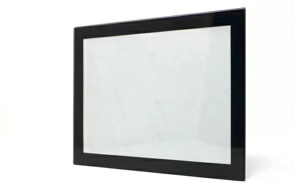
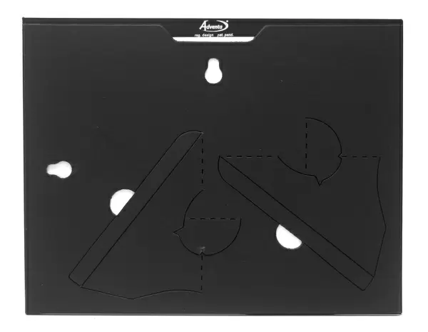
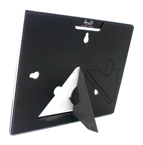
8"x6" Glass Mount
Wall mounted or free-standing, these black edged glass frames feature a smooth chamfered edge and a stylish black border (on back face of the glass). Manufactured from 4mm thick glass, Glass Mounts are a durable, professional way of displaying and protecting your prints. Your 8x6 print is slotted into the back of the frame so can easily be changed if needed.
Tempered Glass Mounts are ideal for wall display, plus the smaller sizes can also be used free-standing via an integral stand
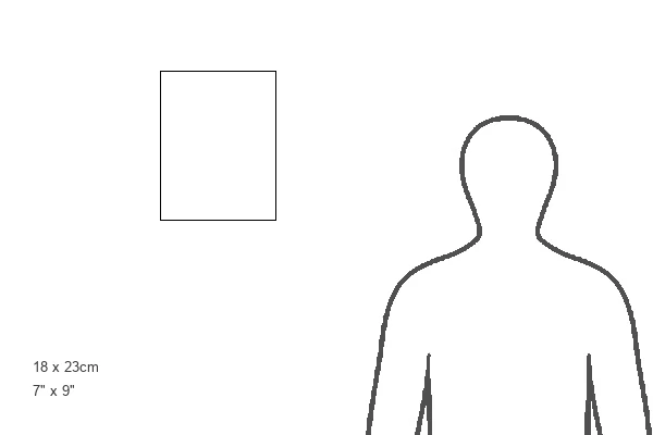
Estimated Image Size (if not cropped) is 15.2cm x 20.3cm (6" x 8")
Estimated Product Size is 17.7cm x 22.8cm (7" x 9")
These are individually made so all sizes are approximate
Artwork printed orientated as per the preview above, with portrait (vertical) orientation to match the source image.
FEATURES IN THESE COLLECTIONS
> Arts
> Minimalist artwork
> Monochrome artwork
> Fine art
> Arts
> Minimalist artwork
> Monochrome artwork
> Monochrome paintings
EDITORS COMMENTS
This print showcases a cross section of human skeletal muscle, revealing its intricate and awe-inspiring structure. The image, taken using transmission electron microscopy (TEM), offers a glimpse into the microscopic world of our muscular system. In this monochrome masterpiece, blocks of skeletal muscle are prominently displayed in lighter grey tones, surrounded by a network of connective tissue represented in black. Delicate filaments composed of actin and myosin proteins can be observed as tiny dots forming the muscle myofibrils. These myofibrils are enclosed by sarcoplasmic reticulum, emphasizing their importance in facilitating muscle contraction. The photograph also highlights the presence of mitochondria - dark grey ovals scattered throughout the image. These organelles play a crucial role in providing the substantial energy required for skeletal muscles to function optimally. It is fascinating to note that skeletal muscles are under conscious control by our brain, enabling us to perform voluntary movements with precision and coordination. This biological marvel exemplifies both health and normalcy within our anatomy. Captured by P. Navarro, R. Bick, and B. Poindexter from UT Medical School/Science Photo Library, this histological masterpiece serves as a testament to the wonders hidden within our bodies' fibers. It invites contemplation on the complexity and beauty inherent in even the smallest units that make up our musculoskeletal system.
MADE IN THE UK
Safe Shipping with 30 Day Money Back Guarantee
FREE PERSONALISATION*
We are proud to offer a range of customisation features including Personalised Captions, Color Filters and Picture Zoom Tools
SECURE PAYMENTS
We happily accept a wide range of payment options so you can pay for the things you need in the way that is most convenient for you
* Options may vary by product and licensing agreement. Zoomed Pictures can be adjusted in the Basket.