Glass Frame > Popular Themes > Human Body
Glass Frame : Immunofluorescent LM of neurons & astrocytes


![]()
Mounted Prints from Science Photo Library
Immunofluorescent LM of neurons & astrocytes
Immunofluorescent Light Micrograph of a network of neurons and astrocyte cells, in brain cortex. In the foreground, nerve fibres of neurons (green) are seen in a fine branching network. These fibres are the routes of transmission of nerve impulses. Astrocytes behind them (orange) are large star- shaped connective tissue cells; as part of the neuroglia (" nerve glue" ) they provide mechanical support and nutrients for the neurons. Astrocytes may take part in information storage processes. Immunofluorescence is a staining technique which uses antibodies to attach fluorescent dyes to specific tissues and to molecules within the cell. Magnification: x200 at 35mm, x375 at 6x4.5cm
Science Photo Library features Science and Medical images including photos and illustrations
Media ID 6421742
© NANCY KEDERSHA/UCLA/SCIENCE PHOTO LIBRARY
Astrocyte Fibres Immunofluores Magnified Image Microscopic Photos Nerve Fibre Nervous Net Work Neuroglia Neurone Subjects System Brain Light Micrograph
7"x5" Glass Mount
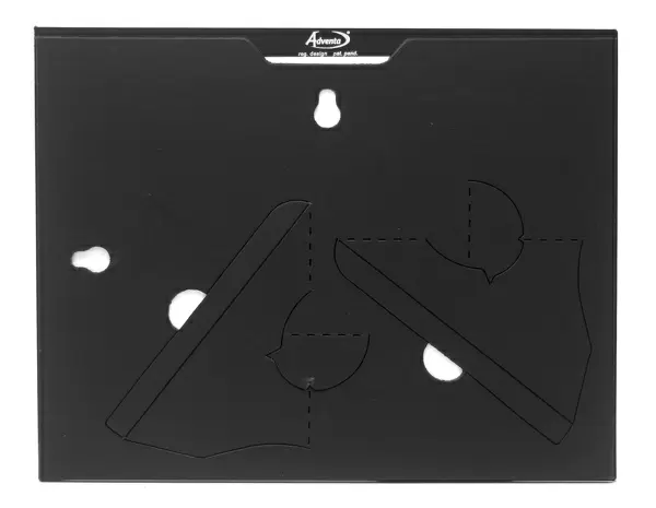
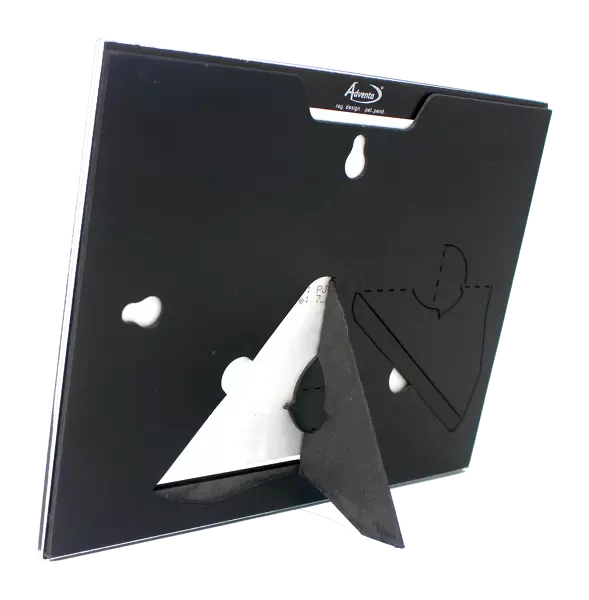
Wall mounted or free-standing, these black edged glass frames feature a smooth chamfered edge and a stylish black border (on back face of the glass). Manufactured from 4mm thick glass, Glass Mounts are a durable, professional way of displaying and protecting your prints. Your 7x5 print is slotted into the back of the frame so can easily be changed if needed.
Tempered Glass Mounts are ideal for wall display, plus the smaller sizes can also be used free-standing via an integral stand
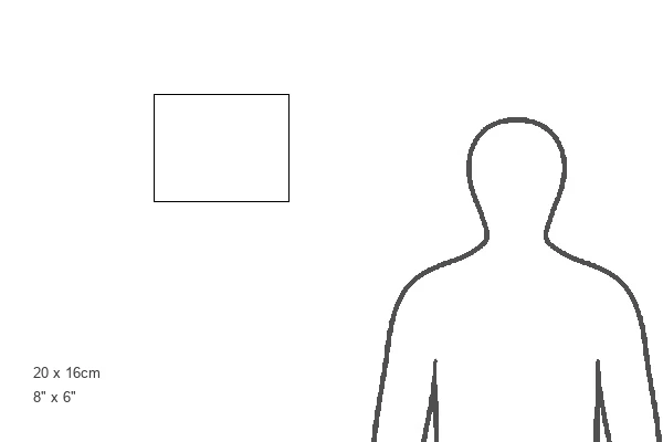
Estimated Image Size (if not cropped) is 17.7cm x 12.7cm (7" x 5")
Estimated Product Size is 20.3cm x 16.2cm (8" x 6.4")
These are individually made so all sizes are approximate
Artwork printed orientated as per the preview above, with landscape (horizontal) orientation to match the source image.
EDITORS COMMENTS
This print showcases the intricate world of neurons and astrocytes in the brain cortex. In a mesmerizing display of colors, nerve fibers of neurons take center stage with their fine branching network, depicted in vibrant green hues. These fibers serve as the vital routes for transmitting nerve impulses throughout the brain. Behind them, large star-shaped connective tissue cells known as astrocytes emerge in striking orange tones. As part of the neuroglia or "nerve glue" these remarkable cells play a crucial role by providing mechanical support and essential nutrients to sustain the neurons. Additionally, there is evidence suggesting that astrocytes may also participate in information storage processes within our complex neural networks. The image was captured using immunofluorescence staining techniques which utilize antibodies to attach fluorescent dyes specifically to tissues and molecules within cells. This technique allows for an enhanced visualization of these microscopic structures. With a magnification level of x200 at 35mm or x375 at 6x4.5cm, this photograph offers us a glimpse into the astonishing complexity and beauty found within our own nervous system. It serves as a reminder of just how intricately interconnected our brains are and highlights the importance of both neurons and astrocytes in maintaining proper brain function.
MADE IN THE UK
Safe Shipping with 30 Day Money Back Guarantee
FREE PERSONALISATION*
We are proud to offer a range of customisation features including Personalised Captions, Color Filters and Picture Zoom Tools
SECURE PAYMENTS
We happily accept a wide range of payment options so you can pay for the things you need in the way that is most convenient for you
* Options may vary by product and licensing agreement. Zoomed Pictures can be adjusted in the Basket.