Glass Frame > Popular Themes > Human Body
Glass Frame : F / col TEM of acinar cells in exocrine pancreas


![]()
Mounted Prints from Science Photo Library
F / col TEM of acinar cells in exocrine pancreas
False-colour transmission electron micrograph (TEM) featuring two elongated, acinar cells of the exocrine human pancreas. Arranged in rounded glands, these cells secrete an alkaline, enzyme rich fluid into the duodenum via the small duct (in blue) at top of image. The pancreatic secretions act to neutralise the acidic contents of the stomach as they move into the small intestine. The pyramidal-shaped cells contain a rounded nucleus (blue) with a prominent nucleoli (red). Granules of pancreatic enzyme pass through the cytoplasm towards the duct at top. Magnification: x3000 at 6x7cm size
Science Photo Library features Science and Medical images including photos and illustrations
Media ID 6450471
© CNRI/SCIENCE PHOTO LIBRARY PHOTO LIBRARY
Acinus Exocrine Pancreas Secretory Cell
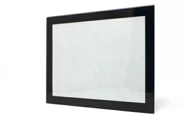
7"x5" Glass Mount
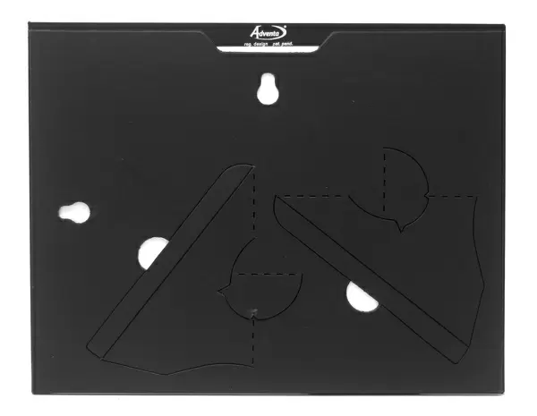
Wall mounted or free-standing, these black edged glass frames feature a smooth chamfered edge and a stylish black border (on back face of the glass). Manufactured from 4mm thick glass, Glass Mounts are a durable, professional way of displaying and protecting your prints. Your 7x5 print is slotted into the back of the frame so can easily be changed if needed.
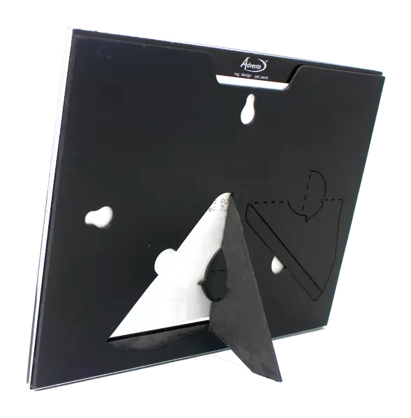
Tempered Glass Mounts are ideal for wall display, plus the smaller sizes can also be used free-standing via an integral stand
Estimated Image Size (if not cropped) is 12.7cm x 17.7cm (5" x 7")
Estimated Product Size is 16.2cm x 20.3cm (6.4" x 8")
These are individually made so all sizes are approximate
Artwork printed orientated as per the preview above, with portrait (vertical) orientation to match the source image.
EDITORS COMMENTS
This print showcases the intricate beauty of acinar cells in the exocrine pancreas. Through false-color transmission electron microscopy, two elongated acinar cells are elegantly displayed within rounded glands. These specialized cells play a vital role in our digestive system by secreting an alkaline, enzyme-rich fluid into the duodenum via a small duct depicted in blue at the top of the image. The pancreatic secretions serve a crucial purpose: neutralizing the acidic contents from our stomach as they enter the small intestine. The pyramidal-shaped acinar cells contain a prominent nucleus portrayed in blue, accompanied by striking nucleoli highlighted in red. As we zoom into this microscopic world, granules of pancreatic enzymes can be observed traversing through the cytoplasm towards their ultimate destination -the duct at the top. At a magnification of x3000 and presented on a 6x7cm size format, this photograph captures not only the mesmerizing structure but also emphasizes its significance within our body's complex machinery. It is truly remarkable how these tiny components work harmoniously to ensure proper digestion and maintain overall health. Science Photo Library has once again provided us with an awe-inspiring glimpse into cellular anatomy and function that leaves us marveling at nature's ingenuity.
MADE IN THE UK
Safe Shipping with 30 Day Money Back Guarantee
FREE PERSONALISATION*
We are proud to offer a range of customisation features including Personalised Captions, Color Filters and Picture Zoom Tools
SECURE PAYMENTS
We happily accept a wide range of payment options so you can pay for the things you need in the way that is most convenient for you
* Options may vary by product and licensing agreement. Zoomed Pictures can be adjusted in the Basket.