
Acinus Collection
Acinus: The Microscopic Marvels of the Body Deep within our bodies lie tiny structures called acini, which play a crucial role in various organs


All Professionally Made to Order for Quick Shipping
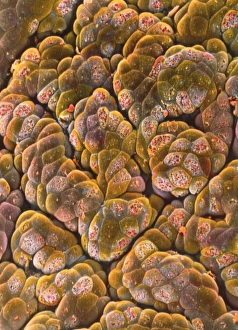
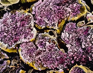
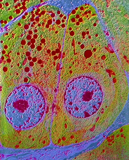
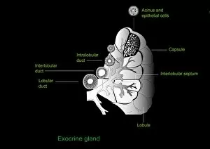
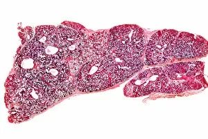
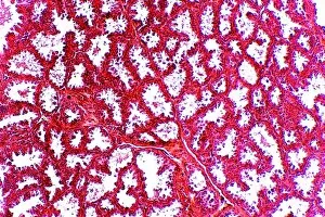
Acinus: The Microscopic Marvels of the Body Deep within our bodies lie tiny structures called acini, which play a crucial role in various organs. One such example is found in the Islet of Langerhans, where these acini cells work tirelessly to regulate blood sugar levels. Captured under a light micrograph, their intricate arrangement showcases the complexity hidden beneath our skin. Moving on to the pancreas, another fascinating sight awaits us - an SEM image revealing the mesmerizing beauty of acini cells. Their unique structure and organization can be observed in this false-color representation that highlights their significance in pancreatic function. Zooming closer into the exocrine pancreas, we delve even deeper with F/col TEM imaging techniques. This high-resolution view provides valuable insights into how acinar cells contribute to digestive processes and enzyme secretion. But let's not forget about our salivary glands. The parotid gland takes center stage as it reveals its own set of acini cells through multiple light micrographs. These snapshots remind us of their vital role in saliva production and maintaining oral health. As we explore further, artwork depicting different types of exocrine glands catches our attention. From sweat glands to mammary glands, each one has its own specialized set of acini cells working diligently behind the scenes. Lastly, we encounter a captivating light micrograph capturing breast tissue during pregnancy. Here again, we witness how acini cells come together to support lactation and nourish newborns. In every corner of our body lies an incredible world waiting to be discovered – from Islets of Langerhans regulating blood sugar levels to parotid salivary glands ensuring proper oral hygiene; from exocrine glands aiding digestion to mammary glands nurturing new life. Acinus serves as a reminder that even at microscopic scales, nature never fails to amaze us with its intricacy and brilliance.