
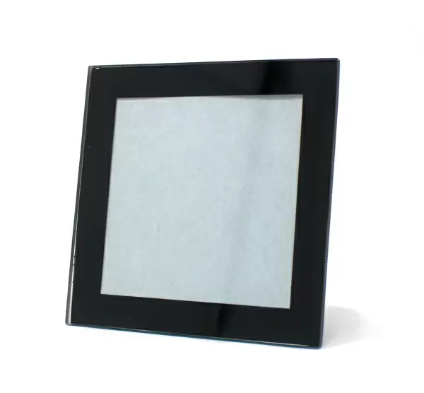
Glass Coaster : Red blood cell crenation, SEM


![]()
Home Decor from Science Photo Library
Red blood cell crenation, SEM
Red blood cell crenation. Coloured scanning electron micrograph (SEM) of two red blood cells (erythrocytes). The cell on the bottom is normal. The cell on the top is crenated (spiked) and is termed an echinocyte. This occurs due to osmosis when a blood sample is exposed to a concentrated solution. It may be symptomatic of uraemia (accumulation of nitrogenous waste in the blood) but more usually is the result of prolonged storage of a blood specimen. Erythrocytes contain the red pigment haemoglobin, allowing them to transport and supply oxygen. Magnification x3250 at 6x7cm size
Science Photo Library features Science and Medical images including photos and illustrations
Media ID 6421049
© STEVE GSCHMEISSNER/SCIENCE PHOTO LIBRARY
Blood Cell Comparison Couple Crenated Crenation Dehydration Echinocyte Erythrocyte Erythrocytes Magnified Image Microscopic Photos Red Blood Cell Spicules Subjects Cells Circulatory System Osmosis
Glass Coaster
Individual Glass Coaster. Stylish and elegant polished safety glass, toughened and heat resistant (10x10cm, 7mm thick). Price shown is per individual coaster.
Individual Glass Coaster. Elegant polished safety toughened glass and heat resistant, matching Place Mats are also available
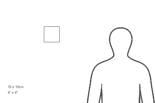
Estimated Image Size (if not cropped) is 7.1cm x 7.6cm (2.8" x 3")
Estimated Product Size is 10cm x 10cm (3.9" x 3.9")
These are individually made so all sizes are approximate
Artwork printed orientated as per the preview above, with portrait (vertical) orientation to match the source image.
EDITORS COMMENTS
This print showcases the intricate details of red blood cells under a scanning electron microscope. The image features two erythrocytes, or red blood cells, with one displaying a normal appearance while the other exhibits crenation, giving it a spiked shape and earning it the name echinocyte. Crenation occurs when a blood sample is exposed to a concentrated solution, causing osmosis to take place. While this phenomenon can be symptomatic of uraemia, which refers to an accumulation of nitrogenous waste in the blood, it is more commonly observed as a result of prolonged storage of a blood specimen. Erythrocytes play a vital role in our circulatory system by containing haemoglobin, the red pigment responsible for transporting and supplying oxygen throughout our bodies. This magnified image at 3250 times its original size allows us to appreciate their delicate structure and understand how dehydration or exposure to hyperosmotic conditions can lead to crenated cells like those depicted here. The contrasting duo presented in this photograph serves as both an educational tool and an artistic representation of cellular biology. Its vibrant colors and meticulous detail make it an intriguing addition to any scientific collection or study on human anatomy.
MADE IN THE UK
Safe Shipping with 30 Day Money Back Guarantee
FREE PERSONALISATION*
We are proud to offer a range of customisation features including Personalised Captions, Color Filters and Picture Zoom Tools
SECURE PAYMENTS
We happily accept a wide range of payment options so you can pay for the things you need in the way that is most convenient for you
* Options may vary by product and licensing agreement. Zoomed Pictures can be adjusted in the Basket.