Fraud Blocker Skip to main content

Membrane Collection (page 9)

The intricate anatomy of the human ear is beautifully depicted in this lithograph, published in 1876

Background imageMembrane Collection: Kidneys, nerves and blood vessels

Kidneys, nerves and blood vessels
Kidneys, historical anatomical artwork. This dorsal (back) view shows the kidneys and associated nerves lying outside the peritoneum (membrane lining the abdominal cavity)

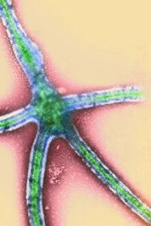
Background imageMembrane Collection: Ebola virus replication, TEM

Ebola virus replication, TEM

Background imageMembrane Collection: Runny nose

Runny nose. Artwork of a section through the ear, nose and throat of a patient with excessive mucus production due to a cold or hay fever (allergic rhinitis)

Background imageMembrane Collection: Ebola virus, TEM

Ebola virus, TEM
Ebola virus. Coloured transmission electron micrograph (TEM) of the extrusion (release) of an Ebola virus (red) from a host cell (green)

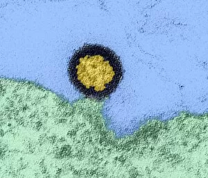
Background imageMembrane Collection: AIDS virus particle, TEM

AIDS virus particle, TEM
AIDS virus particle. Coloured transmission electron micrograph (TEM) of a section through an AIDS virus particle leaving a host cell (green, lower frame)

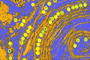
Background imageMembrane Collection: Coloured TEM of herpes simplex viruses inside cell

Coloured TEM of herpes simplex viruses inside cell
Herpes simplex viruses. Coloured transmission electron micrograph (TEM) of a section through a cell infected with herpes simplex viruses

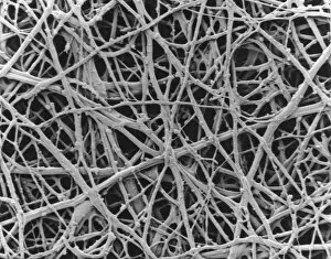
Background imageMembrane Collection: SEM of internal membrane of hen eggshell

SEM of internal membrane of hen eggshell
Eggshell membrane. Scanning electron micrograph (SEM) of the internal membrane from the shell of a hens egg. Several membranes are found around the embryo in a birds egg

Background imageMembrane Collection: Nasal polyp, light micrograph

Nasal polyp, light micrograph
Nasal polyp. Light micrograph of a section through a nasal polyp, a benign growth that arises from a mucous membrane. Magnification: x150 when printed at 10 centimetres wide

Background imageMembrane Collection: Freeze fracture micrograph of cell nucleus

Freeze fracture micrograph of cell nucleus
Cell nucleus. Freeze-fracture scanning electron micrograph of a cell from a parathyroid gland. In the lower left is the nucleus (orange), studded with nuclear pores

Background imageMembrane Collection: Potassium channel molecular model

Potassium channel molecular model
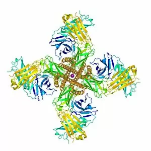
Potassium channel research. Molecular model of the molecular structure of a KcsA potassium ion (K+) channel (brown spirals, centre) from a mouse (mus musculus)

Background imageMembrane Collection: TEM of a cell nucleus membrane showing pores

TEM of a cell nucleus membrane showing pores
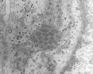
Cell nucleus membrane. Transmission electron micrograph (TEM) of a section through part of the nuclear membrane of a kidney cell revealing its nuclear pores

Background imageMembrane Collection: Simulated space ice

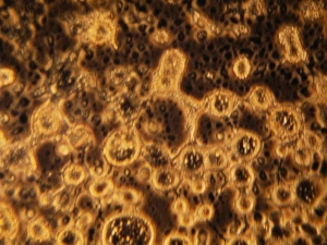
Simulated space ice. Light micrograph of vesicles (round structures) in ice formed under conditions simulating those in space

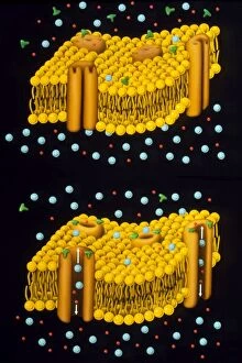
Background imageMembrane Collection: Illustration of ion chanels in plasma membrane

Illustration of ion chanels in plasma membrane
Illustration of a model for the potassium and sodium ion channels through pores in the plasma membrane of an animal nerve cell: an example of active transport, where solutes (ions, sugars)

Background imageMembrane Collection: Col. freeze-fracture TEM of cell nucleus membrane

Col. freeze-fracture TEM of cell nucleus membrane
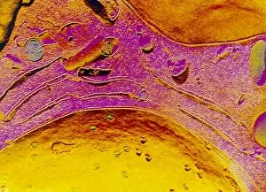
Cell nucleus membrane. Coloured freeze-fracture transmission electron micrograph (TEM) of part of the nuclear membrane of a liver cell

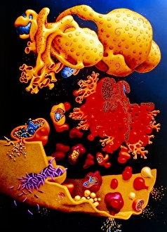
Background imageMembrane Collection: Artwork of ER, golgi body, plasma membrane of cell

Artwork of ER, golgi body, plasma membrane of cell
Illustration of membrane structures in a cell: endoplasmic reticulum, golgi apparatus and plasma membrane. At top, the nucleus is seen with pores, & ribosomes (pink dots) on its surface

Background imageMembrane Collection: Mitochondria

Mitochondria. Coloured transmission electron micrograph of mitochondria (circles). A mitochondrion is a membrane-bound organelle that exists in eukaryotic cells

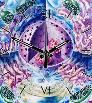
Background imageMembrane Collection: Cellular clock

Cellular clock. Conceptual artwork of a clock and the internal structures (organelles) of an animal cell. This could represent the rate of metabolic cell reactions

Background imageMembrane Collection: Liposomes, SEM

Liposomes, SEM

Background imageMembrane Collection: Evolving protocell, artwork

Evolving protocell, artwork
Evolving protocell. Image 3 of 5. Artwork showing a protocell (artificial cell). This protocell was formed from a synthetic primeval soup containing PNA (peptide nucleic acid, orange)

Background imageMembrane Collection: Liposomes, TEM

Liposomes, TEM

Background imageMembrane Collection: Newly formed protocell, artwork

Newly formed protocell, artwork
Newly formed protocell. Image 2 of 5. Artwork showing cellular components inside a basic cell membrane. This protocell (artificial cell)

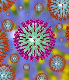
Background imageMembrane Collection: Herpes virus particles, artwork

Herpes virus particles, artwork
Herpes virus particles, computer artwork. Each particle (virion) consists of DNA (deoxyribonucleic acid) contained within a capsid and an envelope covered in glycoprotein spikes

Background imageMembrane Collection: Cell nucleus, TEM

Cell nucleus, TEM
Cell nucleus. Coloured transmission electron micrograph (TEM) of a section through a cell, showing the nucleus (large, spherical), and mitochondria (green)

Background imageMembrane Collection: Cryptochrome and rhodopsin pigments

Cryptochrome and rhodopsin pigments. Rhodopsin is a pigment found in the rod photoreceptor cells in the retina of the human eye, where it absorbs light falling on the retina

Background imageMembrane Collection: Outer membrane receptor protein molecule

Outer membrane receptor protein molecule
Outer membrane receptor protein. Molecular model of FecA an outer membrane receptor protein

Background imageMembrane Collection: Common pheasant male

Common pheasant male
Head of adult male common pheasant showing wattles and displaying the nictitating eye membrane. Dorset, UK March 2011

Background imageMembrane Collection: Cytoskeleton and membrane, diagram

Cytoskeleton and membrane, diagram
Cytoskeleton and membrane. Diagram showing the various structures associated with the cytoskeleton, the protein scaffolding found within cells

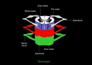
Background imageMembrane Collection: Meninges, artwork

Meninges, artwork
Meninges. Computer artwork showing the meninges surrounding the spinal cord. The meninges are the three membranes that envelop the brain and spinal cord

Background imageMembrane Collection: Immune system, artwork

Immune system, artwork
Immune system. Computer artwork showing how T-cells (right), antibodies (Y-shaped) and antigen-presenting cells (APC, left) interact during an immune response

Background imageMembrane Collection: Perforated eardrum, artwork

Perforated eardrum, artwork
Perforated eardrum. Artwork of a perforated (torn) eardrum (right), or tympanic membrane, and a normal eardrum (left), as seen from the outer ear

Background imageMembrane Collection: Synapse structure, artwork

Synapse structure, artwork
Synapse structure. Cutaway artwork showing the structure of a synapse, the point where two nerve ending meet. The electrical impulse moving along a nerve is transmitted to the adjacent nerve by

Background imageMembrane Collection: Bacterial endospore formation, diagram

Bacterial endospore formation, diagram. The initial stage is at upper left, following the arrow to a ninth stage at lower right

Background imageMembrane Collection: Chloroplast structures, artwork

Chloroplast structures, artwork
Chloroplast structures. Cutaway artwork showing the internal structure of a chloroplast, the organelle in plant cells responsible for photosynthesis

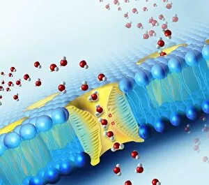
Background imageMembrane Collection: Aquaporins, artwork

Aquaporins, artwork
Aquaporins. Computer artwork of water molecules (red and white) passing through pores (yellow) in a cell membrane (blue). These pores are proteins known as aquaporins that are embedded in

Background imageMembrane Collection: Aquaporin roles in the body

Aquaporin roles in the body. Aquaporins are membrane proteins that form channels (lower right) that help water molecules pass in and out of cells

Background imageMembrane Collection: Mitochondrion, artwork

Mitochondrion, artwork
Mitochondrion. Cutaway artwork showing the internal structure of a mitochondrion. This structure, found in eukaryotic cells, is the site of energy production

Background imageMembrane Collection: Animal cell processes, artwork

Animal cell processes, artwork
Animal cell processes. Cutaway artwork showing the structures inside an animal cell and four different processes that take place inside it or on its membrane (all marked by magnifying glasses)

Background imageMembrane Collection: Animal cell structure, artwork

Animal cell structure, artwork
Animal cell structure. Artwork showing the internal structure of an animal cell. Inside the cell, the cellular structures (organelles) include the Golgi apparatus (green)

Background imageMembrane Collection: Multiverse layers, artwork

Multiverse layers, artwork. The multiverse, also called the meta-universe or metaverse, is the hypothetical set of possible universes, and includes all of space and time

Background imageMembrane Collection: Golgi apparatus, artwork

Golgi apparatus, artwork. This structure is an organelle found within eukaryotic cells. It receives proteins and lipids that are synthesised elsewhere on the endoplasmic reticulum

Background imageMembrane Collection: Desalination Reverse Osmosis filter

Desalination Reverse Osmosis filter
Desalination plant. A bank of Reverse Osmosis membrane filters. This facility turns salt water into drinking water using the Reverse Osmosis Process

Background imageMembrane Collection: Bacterial cell structure, artwork

Bacterial cell structure, artwork
Bacterial cell structure. Computer artwork showing the cell structure and components (organelles) of a typical rod-shaped bacteria (bacillus). Not all bacteria have a flagellum (long, tail-like)

Background imageMembrane Collection: Bacterial cell wall comparison, artwork

Bacterial cell wall comparison, artwork
Bacterial cell wall comparison. Computer artwork comparing the structure of the cell wall from a gram-negative bacterium (left) with that of a gram-positive bacterium (right)

Background imageMembrane Collection: Domestic greenhouse under construction

Domestic greenhouse under construction. The greenhouse structure is mounted on a base, complete with weed control fabric, but is unglazed

Background imageMembrane Collection: Domestic greenhouse in garden

Domestic greenhouse in garden
Domestic greenhouse under construction. The greenhouse is glazed and the base is lined with weed control fabric. There is no staging

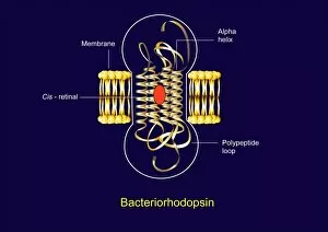
Background imageMembrane Collection: Bacteriorhodopsin, diagram

Bacteriorhodopsin, diagram
Bacteriorhodopsin. Diagram of the molecular structure of bacteriorhodopsin, a protein found in primitive micro-organisms known as Archaea

Background imageMembrane Collection: Bacteriorhodopsin, artwork

Bacteriorhodopsin, artwork
Bacteriorhodopsin. Artwork of the molecular structure of bacteriorhodopsin, a protein found in primitive micro-organisms known as Archaea

Background imageMembrane Collection: Muscle anatomy

Muscle anatomy. Cutaway artwork showing the anatomy and internal structure of a muscle, from the macroscopic (left) to the microscopic (right) level


All Professionally Made to Order for Quick Shipping

The intricate anatomy of the human ear is beautifully depicted in this lithograph, published in 1876. One notable feature highlighted here is the membrane, which plays a crucial role in transmitting sound waves to our auditory system. Moving on to cellular structures, the rough endoplasmic reticulum (ER) takes center stage under a transmission electron microscope (TEM). This network of membranes within cells is responsible for protein synthesis and transport. Artwork showcasing different cell types also emphasizes the significance of membranes. From the delicate cell membrane itself, represented by artwork C013 / 7467, to mitochondria seen through TEM imaging - these organelles possess their own unique membranes that regulate various cellular functions. Intriguingly, even chloroplasts have their own distinct membrane structure as revealed by artwork dedicated to studying photosynthesis. These specialized organelles found in plants are responsible for converting sunlight into energy. Beyond biology, membranes find relevance elsewhere too. Think about damp-proofing measures taken in houses – membranes act as barriers against moisture infiltration and protect our living spaces from potential damage. However, not all mentions of they are positive. Bacterial meningitis can be detected through MRI scans where inflammation affects the protective brain meninges' integrity. Understanding how pathogens breach these defensive layers helps diagnose and treat such infections effectively. Nature's wonders also exhibit fascinating adaptations involving membranes; take Plecotus sp. , commonly known as long-eared bats with their remarkable hearing abilities thanks to specialized ear membrane structures aiding echolocation skills. Zooming into finer details under TEM again reveals eye muscles' intricate arrangement (TEM C014 / 1468), highlighting how well-organized muscle fibers rely on precise membranous connections for coordinated movement and vision control. Lastly, let's not forget intestinal microvilli observed through TEM – finger-like projections covered by plasma membrane lining our intestines play a vital role in nutrient absorption during digestion processes. From ancient lithographs to modern imaging techniques, the significance of membranes spans across various fields.