Fraud Blocker Skip to main content

Membrane Collection (page 5)

The intricate anatomy of the human ear is beautifully depicted in this lithograph, published in 1876

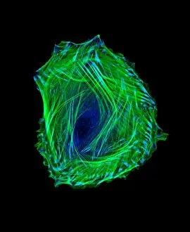
Background imageMembrane Collection: Embryonic smooth muscle cell C018 / 8595

Embryonic smooth muscle cell C018 / 8595
Embryonic smooth muscle cell, immunofluorescence micrograph. Actin filaments are green, the protein vinculin is light blue. Actin is part of the cytoskeleton

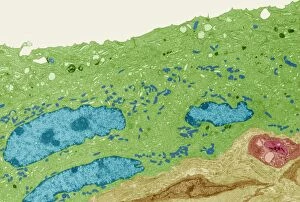
Background imageMembrane Collection: Bladder lining, TEM C014 / 1473

Bladder lining, TEM C014 / 1473
Bladder lining. Transmission electron micrograph (TEM) of a section through the epithelial cells that line of a human bladder, showing large flat vesicles (large, blue)

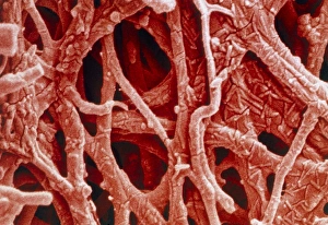
Background imageMembrane Collection: Coloured SEM of internal membrane of hen eggshell

Coloured SEM of internal membrane of hen eggshell
Eggshell membrane. Coloured scanning electron micrograph (SEM) of the internal membrane from the shell of a hens egg. Several membranes are found around the embryo in a birds egg

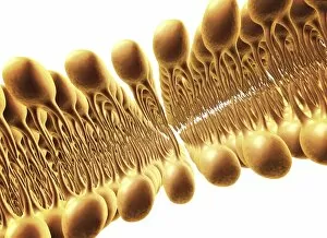
Background imageMembrane Collection: Cell membrane lipid bilayer, artwork F007 / 1479

Cell membrane lipid bilayer, artwork F007 / 1479
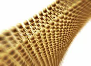
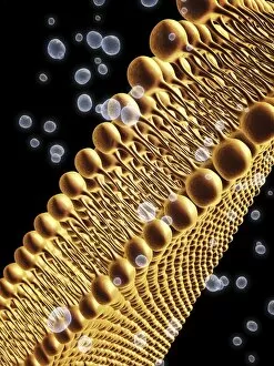
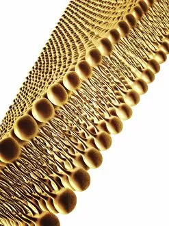
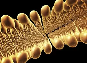
Phospholipid bilayer. Computer artwork of the phospholipid bilayer that forms the membrane around all living cells. The cell membrane is made of phospholipid molecules

Background imageMembrane Collection: Cell membrane lipid bilayer, artwork F007 / 1480

Cell membrane lipid bilayer, artwork F007 / 1480
Phospholipid bilayer. Computer artwork of the phospholipid bilayer that forms the membrane around all living cells. The cell membrane is made of phospholipid molecules

Background imageMembrane Collection: Cell membrane lipid bilayer, artwork F007 / 1478

Cell membrane lipid bilayer, artwork F007 / 1478
Phospholipid bilayer. Computer artwork of the phospholipid bilayer that forms the membrane around all living cells. The cell membrane is made of phospholipid molecules

Background imageMembrane Collection: Cell membrane lipid bilayer, artwork F007 / 1475

Cell membrane lipid bilayer, artwork F007 / 1475
Phospholipid bilayer. Computer artwork of the phospholipid bilayer that forms the membrane around all living cells. The cell membrane is made of phospholipid molecules

Background imageMembrane Collection: Cell membrane lipid bilayer, artwork F007 / 1474

Cell membrane lipid bilayer, artwork F007 / 1474
Phospholipid bilayer. Computer artwork of the phospholipid bilayer that forms the membrane around all living cells. The cell membrane is made of phospholipid molecules

Background imageMembrane Collection: Cell membrane lipid bilayer, artwork F007 / 1473

Cell membrane lipid bilayer, artwork F007 / 1473
Phospholipid bilayer. Computer artwork of the phospholipid bilayer that forms the membrane around all living cells. The cell membrane is made of phospholipid molecules

Background imageMembrane Collection: Wnt signalling pathways, illustration C018 / 0917

Wnt signalling pathways, illustration C018 / 0917
Wnt signalling pathways, illustration. Wnt signalling pathways are three separate pathways that pass signals from outside a cell to inside the cell

Background imageMembrane Collection: Nucleus and endoplasmic reticulum F006 / 9196

Nucleus and endoplasmic reticulum F006 / 9196
Computer artwork showing part of a human or eukaryotic cell. In the middle the nucleus which has a membrane with nuclear pores. Inside the nucleus is the DNA

Background imageMembrane Collection: Palmitoyloleoyl PE lipid bilayer F006 / 9780

Palmitoyloleoyl PE lipid bilayer F006 / 9780
Palmitoyloleoyl PE (POPE) lipid bilayer, computer simulation. This is a model of the phospholipid bilayer of the inner bacterial membrane

Background imageMembrane Collection: Outer membrane receptor protein molecule F006 / 9398

Outer membrane receptor protein molecule F006 / 9398
Outer membrane receptor protein. Molecular model of FecA an outer membrane receptor protein

Background imageMembrane Collection: Sucrose-specific porin molecule F006 / 9218

Sucrose-specific porin molecule F006 / 9218
Sucrose-specific porin, molecular model. Porins are proteins that span cell membranes and act as a channel through which specific molecules can diffuse

Background imageMembrane Collection: Nucleus and endoplasmic reticulum F006 / 9201

Nucleus and endoplasmic reticulum F006 / 9201
Computer artwork showing part of a human or eukaryotic cell. In the middle the nucleus which has a membrane with nuclear pores. Inside the nucleus is the DNA

Background imageMembrane Collection: Thin-film thermocouples C016 / 6482

Thin-film thermocouples C016 / 6482
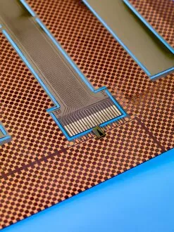
Thin-film thermocouples. Thermocouples, used for temperature measurements, are based on the thermoelectric effect between two metals

Background imageMembrane Collection: Voice box anatomy, artwork C016 / 7692

Voice box anatomy, artwork C016 / 7692
Voice box anatomy. Computer artwork showing the location (right) and structure (left) of a human larynx (voice box). The larynx is involved in breathing, sound production (phonation)

Background imageMembrane Collection: E. coli bacteria, SEM C016 / 9128

E. coli bacteria, SEM C016 / 9128
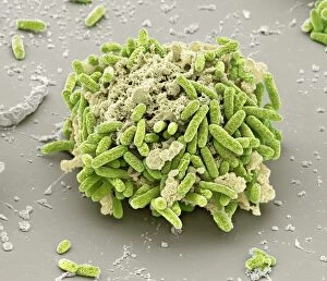
E. coli bacteria. Coloured scanning electron micrograph (SEM) of Escherichia coli bacteria (purple) attached to the remains of a dead cell. E

Background imageMembrane Collection: Shreves Sarayacu treefrog calling

Shreves Sarayacu treefrog calling. Shreves Sarayacu treefrog (Dendropsophus sarayacuensis) is found in Bolivia, Brazil, Colombia, Ecuador, Peru, and Venezuela

Background imageMembrane Collection: E. coli bacteria, SEM C016 / 9130

E. coli bacteria, SEM C016 / 9130
E. coli bacteria. Coloured scanning electron micrograph (SEM) of Escherichia coli bacteria (red) attached to the remains of a dead cell. E

Background imageMembrane Collection: Thin-film thermocouple C016 / 6484

Thin-film thermocouple C016 / 6484
Thin-film thermocouple, close-up. Thermocouples, used for temperature measurements, are based on the thermoelectric effect between two metals

Background imageMembrane Collection: E. coli bacteria, SEM C016 / 9127

E. coli bacteria, SEM C016 / 9127
E. coli bacteria. Coloured scanning electron micrograph (SEM) of Escherichia coli bacteria (green) attached to the remains of a dead cell. E

Background imageMembrane Collection: Thin-film thermocouple C016 / 6486

Thin-film thermocouple C016 / 6486
Thin-film thermocouple, close-up. Thermocouples, used for temperature measurements, are based on the thermoelectric effect between two metals

Background imageMembrane Collection: Thin-film thermocouple C016 / 6483

Thin-film thermocouple C016 / 6483
Thin-film thermocouple. Thermocouples, used for temperature measurements, are based on the thermoelectric effect between two metals

Background imageMembrane Collection: Thin-film thermocouple C016 / 6485

Thin-film thermocouple C016 / 6485
Thin-film thermocouple, close-up. Thermocouples, used for temperature measurements, are based on the thermoelectric effect between two metals

Background imageMembrane Collection: Broken shoulder, artwork C016 / 6568

Broken shoulder, artwork C016 / 6568
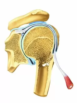
Broken shoulder. Artwork of a vertical (coronal) section through the bones of the shoulder, showing a fracture (centre right) of the upper part of the humerus (upper arm bone)

Background imageMembrane Collection: Thin-film thermocouples C016 / 6481

Thin-film thermocouples C016 / 6481
Thin-film thermocouples. Thermocouples, used for temperature measurements, are based on the thermoelectric effect between two metals

Background imageMembrane Collection: Thin-film thermocouples C016 / 6480

Thin-film thermocouples C016 / 6480
Thin-film thermocouples. Thermocouples, used for temperature measurements, are based on the thermoelectric effect between two metals

Background imageMembrane Collection: Cranium anatomy, artwork

Cranium anatomy, artwork
Cranium anatomy, computer artwork. At bottom is the brain, which is enclosed in three membranes (grey) known as the meninges

Background imageMembrane Collection: Smell receptor, TEM

Smell receptor, TEM
Smell receptor. Transmission electron micrograph (TEM) of a section through the olfactory epithelium that lines the nasal cavity, showing an olfactory cell (smell receptor)

Background imageMembrane Collection: Golgi membranes, TEM

Golgi membranes, TEM
Golgi membranes. Transmission electron micrograph (TEM) of a section through a cell, showing the membranes (dark lines) of the Golgi apparatus

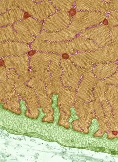
Background imageMembrane Collection: Myelinated nerve tissue, TEM

Myelinated nerve tissue, TEM
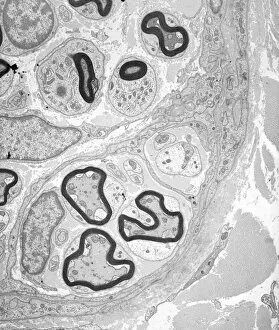
Myelinated nerve tissue. Transmission electron micrograph (TEM) of a section through myelinated nerve fibres (axons). Each axon is coated with many layers of myelin

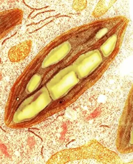
Background imageMembrane Collection: Chloroplast, TEM C016 / 6297

Chloroplast, TEM C016 / 6297
Chloroplast. Coloured transmission electron micrograph (TEM) of chloroplast from the moss Physcomitrella patens. Chloroplasts are the sites of photosynthesis

Background imageMembrane Collection: Eukaryote cell, artwork C016 / 6260

Eukaryote cell, artwork C016 / 6260
Eukaryote cell. Computer artwork showing the internal structure of a typical eukaryotic cell. Eukaryotes are organisms whose cells contain a membrane-bound nucleus (karyon, orange)

Background imageMembrane Collection: Chloroplast, TEM C016 / 6298

Chloroplast, TEM C016 / 6298
Chloroplast. Coloured transmission electron micrograph (TEM) of chloroplast from the moss Physcomitrella patens. Chloroplasts are the sites of photosynthesis

Background imageMembrane Collection: Influenza virus, illustration C018 / 0735

Influenza virus, illustration C018 / 0735
Influenza virus. Illustration of an influenza (flu) virus particle (virion). The virus consists of an RNA (ribonucleic acid) core (black)

Background imageMembrane Collection: Sperm cell, artwork C018 / 6996

Sperm cell, artwork C018 / 6996
Sperm cell anatomy. Cutaway computer artwork showing the internal structure of a sperm cell (spermatozoon), the male sex cell

Background imageMembrane Collection: Eye muscle, TEM C014 / 1467

Eye muscle, TEM C014 / 1467
Eye muscle. Transmission electron micrograph (TEM) of a section through a striated muscle cell from the ciliary muscle of a human eye

Background imageMembrane Collection: Eye muscle, TEM C014 / 1466

Eye muscle, TEM C014 / 1466
Eye muscle. Transmission electron micrograph (TEM) of a section through a striated muscle cell from the ciliary muscle of a human eye

Background imageMembrane Collection: Coagulation factor complex molecule C014 / 0409

Coagulation factor complex molecule C014 / 0409
Coagulation factor complex molecule. Molecular model showing the interaction between coagulation factor VIII (FVIII, pink, blue and yellow), factor IXa (FIXa)

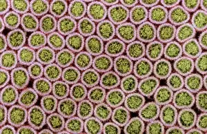
Background imageMembrane Collection: Intestinal microvilli, SEM C014 / 1452

Intestinal microvilli, SEM C014 / 1452
Intestinal microvilli. Coloured transmission electron micrograph (SEM) of a transverse section through microvilli, showing their interiors

Background imageMembrane Collection: Intestinal microvilli, SEM C014 / 1451

Intestinal microvilli, SEM C014 / 1451
Intestinal microvilli. Coloured transmission electron micrograph (SEM) of a transverse section through microvilli, showing their interiors

Background imageMembrane Collection: Coagulation factor complex molecule C014 / 0410

Coagulation factor complex molecule C014 / 0410
Coagulation factor complex molecule. Molecular model showing the interaction between coagulation factor VIII (FVIII, pink, blue and yellow), factor IXa (FIXa)

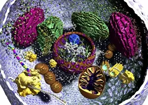
Background imageMembrane Collection: Animal cell organelles, artwork

Animal cell organelles, artwork
Animal cell organelles. Artwork showing the organelles in a eukaryotic cell. This is an animal cell. Structures include the nucleus (centre) which has a membrane with nuclear pores (purple)

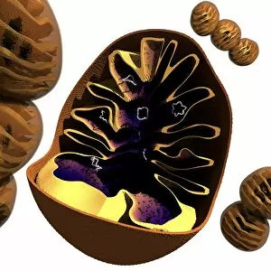
Background imageMembrane Collection: Mitochondrion structure, artwork

Mitochondrion structure, artwork
Mitochondrion structure. Artwork showing the internal structure of a mitochondrion. This structure, found in eukaryotic cells, is the site of energy production

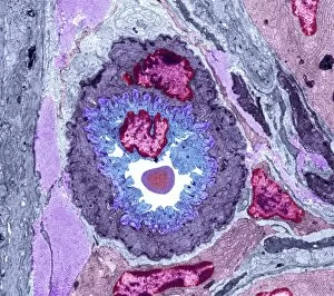
Background imageMembrane Collection: Intestinal arteriole, TEM

Intestinal arteriole, TEM
Intestinal arteriole. Transmission electron micrograph (TEM) of a section through an arteriole in the wall of the small intestine. Magnification: x5000 when printed 10 centimetres wide

Background imageMembrane Collection: Muscle arteriole, TEM

Muscle arteriole, TEM
Muscle arteriole. Transmission electron micrograph (TEM) of a section through an arteriole (black, centre) in striated muscle tissue. Magnification: x3500 when printed 10 centimetres wide

Background imageMembrane Collection: Mitochondrial structure, artwork

Mitochondrial structure, artwork
Mitochondrial structure. Computer artwork of a section through a mitochondrion, showing the internal structure and a loop of mitochondrial DNA (deoxyribonucleic acid, mtDNA)


All Professionally Made to Order for Quick Shipping

The intricate anatomy of the human ear is beautifully depicted in this lithograph, published in 1876. One notable feature highlighted here is the membrane, which plays a crucial role in transmitting sound waves to our auditory system. Moving on to cellular structures, the rough endoplasmic reticulum (ER) takes center stage under a transmission electron microscope (TEM). This network of membranes within cells is responsible for protein synthesis and transport. Artwork showcasing different cell types also emphasizes the significance of membranes. From the delicate cell membrane itself, represented by artwork C013 / 7467, to mitochondria seen through TEM imaging - these organelles possess their own unique membranes that regulate various cellular functions. Intriguingly, even chloroplasts have their own distinct membrane structure as revealed by artwork dedicated to studying photosynthesis. These specialized organelles found in plants are responsible for converting sunlight into energy. Beyond biology, membranes find relevance elsewhere too. Think about damp-proofing measures taken in houses – membranes act as barriers against moisture infiltration and protect our living spaces from potential damage. However, not all mentions of they are positive. Bacterial meningitis can be detected through MRI scans where inflammation affects the protective brain meninges' integrity. Understanding how pathogens breach these defensive layers helps diagnose and treat such infections effectively. Nature's wonders also exhibit fascinating adaptations involving membranes; take Plecotus sp. , commonly known as long-eared bats with their remarkable hearing abilities thanks to specialized ear membrane structures aiding echolocation skills. Zooming into finer details under TEM again reveals eye muscles' intricate arrangement (TEM C014 / 1468), highlighting how well-organized muscle fibers rely on precise membranous connections for coordinated movement and vision control. Lastly, let's not forget intestinal microvilli observed through TEM – finger-like projections covered by plasma membrane lining our intestines play a vital role in nutrient absorption during digestion processes. From ancient lithographs to modern imaging techniques, the significance of membranes spans across various fields.