Fraud Blocker Skip to main content

Membrane Collection (page 6)

The intricate anatomy of the human ear is beautifully depicted in this lithograph, published in 1876

Background imageMembrane Collection: Coagulation factor complex molecule C014 / 0410

Coagulation factor complex molecule C014 / 0410
Coagulation factor complex molecule. Molecular model showing the interaction between coagulation factor VIII (FVIII, pink, blue and yellow), factor IXa (FIXa)

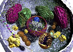
Background imageMembrane Collection: Animal cell organelles, artwork

Animal cell organelles, artwork
Animal cell organelles. Artwork showing the organelles in a eukaryotic cell. This is an animal cell. Structures include the nucleus (centre) which has a membrane with nuclear pores (purple)

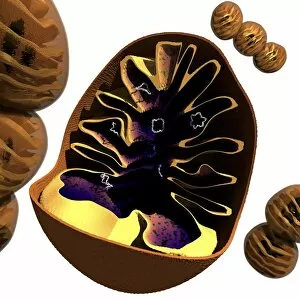
Background imageMembrane Collection: Mitochondrion structure, artwork

Mitochondrion structure, artwork
Mitochondrion structure. Artwork showing the internal structure of a mitochondrion. This structure, found in eukaryotic cells, is the site of energy production

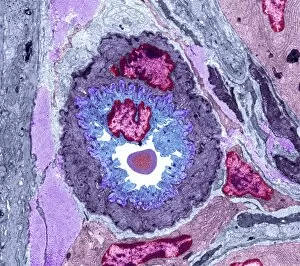
Background imageMembrane Collection: Intestinal arteriole, TEM

Intestinal arteriole, TEM
Intestinal arteriole. Transmission electron micrograph (TEM) of a section through an arteriole in the wall of the small intestine. Magnification: x5000 when printed 10 centimetres wide

Background imageMembrane Collection: Muscle arteriole, TEM

Muscle arteriole, TEM
Muscle arteriole. Transmission electron micrograph (TEM) of a section through an arteriole (black, centre) in striated muscle tissue. Magnification: x3500 when printed 10 centimetres wide

Background imageMembrane Collection: Mitochondrial structure, artwork

Mitochondrial structure, artwork
Mitochondrial structure. Computer artwork of a section through a mitochondrion, showing the internal structure and a loop of mitochondrial DNA (deoxyribonucleic acid, mtDNA)

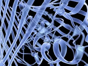
Background imageMembrane Collection: Bacterial outer membrane protein molecule C014 / 4949

Bacterial outer membrane protein molecule C014 / 4949
Bacterial outer membrane protein molecule. Computer model showing the secondary structure of a molecule of outer membrane transporter FecA protein from Escherichia coli (E. coli) bacteria

Background imageMembrane Collection: Bacterial outer membrane protein molecule

Bacterial outer membrane protein molecule. Computer model showing a part of the secondary structure of a molecule of outer membrane protein A from Escherichia coli (E. coli) bacteria

Background imageMembrane Collection: Smooth endoplasmic reticulum, TEM

Smooth endoplasmic reticulum, TEM
Smooth endoplasmic reticulum. Transmission electron micrograph (TEM) showing smooth endoplasmic reticulum (ER, thin lines) inside a cell that is synthesising steroid hormones

Background imageMembrane Collection: Small bowel epithelium, TEM

Small bowel epithelium, TEM
Small bowel epithelium. Transmission electron micrograph (TEM) of a section through the epithelial lining of a villus in the small bowel, showing the enterocyte cells

Background imageMembrane Collection: Influenza virus, artwork C016 / 8349

Influenza virus, artwork C016 / 8349
Influenza virus. Cut-away computer artwork of an influenza (flu) virus particle (virion). In each particles lipid envelope (green) are two types of protein spike, haemagglutinin (H)

Background imageMembrane Collection: Influenza virus, artwork C016 / 8348

Influenza virus, artwork C016 / 8348
Influenza virus. Cut-away computer artwork of an influenza (flu) virus particle (virion). In each particles lipid envelope (green) are two types of protein spike, haemagglutinin (H)

Background imageMembrane Collection: Influenza virus, artwork C016 / 8347

Influenza virus, artwork C016 / 8347
Influenza virus. Cut-away computer artwork of an influenza (flu) virus particle (virion). In each particles lipid envelope (green) are two types of protein spike, haemagglutinin (H)

Background imageMembrane Collection: Influenza virus, artwork C016 / 8346

Influenza virus, artwork C016 / 8346
Influenza virus. Cut-away computer artwork of an influenza (flu) virus particle (virion). In each particles lipid envelope (green) are two types of protein spike, haemagglutinin (H)

Background imageMembrane Collection: Influenza virus, artwork C016 / 8344

Influenza virus, artwork C016 / 8344
Influenza virus. Cut-away computer artwork of an influenza (flu) virus particle (virion). In each particles lipid envelope (green) are two types of protein spike, haemagglutinin (H)

Background imageMembrane Collection: Influenza virus, artwork C016 / 8345

Influenza virus, artwork C016 / 8345
This image may not be used in educational posters Influenza virus. Cut-away computer artwork of an influenza (flu) virus particle (virion)

Background imageMembrane Collection: Influenza virus, artwork C016 / 8342

Influenza virus, artwork C016 / 8342
Influenza virus. Computer artwork of an influenza (flu) virus particle (virion). In each particles lipid envelope (green) are two types of protein spike, haemagglutinin (H, red) and neuraminidase (N)

Background imageMembrane Collection: Influenza virus, artwork C016 / 8341

Influenza virus, artwork C016 / 8341
Influenza virus. Computer artwork of an influenza (flu) virus particle (virion). In each particles lipid envelope (green) are two types of protein spike, haemagglutinin (H, red) and neuraminidase (N)

Background imageMembrane Collection: Influenza virus, artwork C016 / 8340

Influenza virus, artwork C016 / 8340
Influenza virus. Computer artwork of an influenza (flu) virus particle (virion). In each particles lipid envelope (green) are two types of protein spike, haemagglutinin (H, red) and neuraminidase (N)

Background imageMembrane Collection: HIV invading white blood cell, artwork

HIV invading white blood cell, artwork
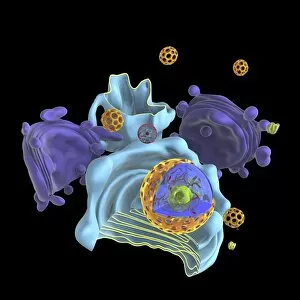
HIV invading white blood cell. Computer artwork showing HIV (human immunodeficiency virus) particles (virions, purple) invading a white blood cell (large, centre) in the human bloodstream

Background imageMembrane Collection: Nucleus and endoplasmic reticulum C015 / 6797

Nucleus and endoplasmic reticulum C015 / 6797
Computer artwork showing part of a human or eukaryotic cell. In the middle the nucleus which has a membrane with nuclear pores. Inside the nucleus is the DNA

Background imageMembrane Collection: Animal cell organelles, artwork C015 / 6793

Animal cell organelles, artwork C015 / 6793
Animal cell organelles. Computer artwork showing the organelles in a eukaryotic cell. This is an animal cell. Structures include the nucleus (centre) which has a membrane with nuclear pores

Background imageMembrane Collection: Animal cell organelles, artwork C015 / 6792

Animal cell organelles, artwork C015 / 6792
Animal cell organelles. Computer artwork showing the organelles in a eukaryotic cell. This is an animal cell. Structures include the nucleus (centre) which has a membrane with nuclear pores

Background imageMembrane Collection: Clathrin lattice, molecular model C015 / 6788

Clathrin lattice, molecular model C015 / 6788
Molecular model of a complete clathrin lattice. The polyhedral protein lattice coats eukaryotic cell membranes (vesicles) and coated pits and appears to be involved in protein secretion

Background imageMembrane Collection: Clathrin lattice, molecular model C015 / 6790

Clathrin lattice, molecular model C015 / 6790
Molecular model of a complete clathrin lattice. The polyhedral protein lattice coats eukaryotic cell membranes (vesicles) and coated pits and appears to be involved in protein secretion

Background imageMembrane Collection: Nucleus and endoplasmic reticulum C015 / 6783

Nucleus and endoplasmic reticulum C015 / 6783
Computer artwork showing part of a human or eukaryotic cell. In the middle the nucleus which has a membrane with nuclear pores. Inside the nucleus is the DNA

Background imageMembrane Collection: Nucleus and endoplasmic reticulum C015 / 6782

Nucleus and endoplasmic reticulum C015 / 6782
Computer artwork showing part of a human or eukaryotic cell. In the middle the nucleus which has a membrane with nuclear pores. Inside the nucleus is the DNA

Background imageMembrane Collection: Nucleus and endoplasmic reticulum C015 / 6781

Nucleus and endoplasmic reticulum C015 / 6781
Computer artwork showing part of a human or eukaryotic cell. In the middle the nucleus which has a membrane with nuclear pores. Inside the nucleus is the DNA

Background imageMembrane Collection: Animal cell organelles, artwork C015 / 6775

Animal cell organelles, artwork C015 / 6775
Animal cell organelles. Computer artwork showing the organelles in a eukaryotic cell. This is an animal cell. Structures include the nucleus (centre) which has a membrane with nuclear pores

Background imageMembrane Collection: Animal cell organelles, artwork C015 / 6778

Animal cell organelles, artwork C015 / 6778
Animal cell organelles. Computer artwork showing the organelles in a eukaryotic cell. This is an animal cell. Structures include the nucleus (centre) which has a membrane with nuclear pores

Background imageMembrane Collection: Animal cell organelles, artwork C015 / 6777

Animal cell organelles, artwork C015 / 6777
Animal cell organelles. Computer artwork showing the organelles in a eukaryotic cell. This is an animal cell. Structures include the nucleus (centre) which has a membrane with nuclear pores

Background imageMembrane Collection: Nucleus and endoplasmic reticulum C015 / 6772

Nucleus and endoplasmic reticulum C015 / 6772
Computer artwork showing part of a human or eukaryotic cell. In the middle the nucleus which has a membrane with nuclear pores. Inside the nucleus is the DNA

Background imageMembrane Collection: Clathrin lattice, molecular model C015 / 6770

Clathrin lattice, molecular model C015 / 6770
Molecular model of a complete clathrin lattice. The polyhedral protein lattice coats eukaryotic cell membranes (vesicles) and coated pits and appears to be involved in protein secretion

Background imageMembrane Collection: Nucleus and endoplasmic reticulum C015 / 6768

Nucleus and endoplasmic reticulum C015 / 6768
Computer artwork showing part of a human or eukaryotic cell. In the middle the nucleus which has a membrane with nuclear pores. Inside the nucleus is the DNA

Background imageMembrane Collection: Nucleus and endoplasmic reticulum C015 / 6767

Nucleus and endoplasmic reticulum C015 / 6767
Computer artwork showing part of a human or eukaryotic cell. In the middle the nucleus which has a membrane with nuclear pores. Inside the nucleus is the DNA

Background imageMembrane Collection: Animal cell organelles, artwork C016 / 0619

Animal cell organelles, artwork C016 / 0619
Computer artwork showing the organelles in a eukaryotic cell. This is an animal cell. Structures include the nucleus (centre, red) which has a membrane with nuclear pores

Background imageMembrane Collection: Animal cell organelles, artwork C016 / 0621

Animal cell organelles, artwork C016 / 0621
Computer artwork showing the organelles in a eukaryotic cell. This is an animal cell. Structures include the nucleus (centre, red) which has a membrane with nuclear pores

Background imageMembrane Collection: Animal cell organelles, artwork C016 / 0620

Animal cell organelles, artwork C016 / 0620
Computer artwork showing the organelles in a eukaryotic cell. This is an animal cell. Structures include the nucleus (centre, orange) which has a membrane with nuclear pores

Background imageMembrane Collection: Animal cell organelles, artwork C016 / 0617

Animal cell organelles, artwork C016 / 0617
Computer artwork showing the organelles in a eukaryotic cell. This is an animal cell. Structures include the nucleus (centre, red) which has a membrane with nuclear pores

Background imageMembrane Collection: Animal cell organelles, artwork C016 / 0618

Animal cell organelles, artwork C016 / 0618
Computer artwork showing the organelles in a eukaryotic cell. This is an animal cell. Structures include the nucleus (centre, orange) which has a membrane with nuclear pores

Background imageMembrane Collection: Animal cell organelles, artwork C016 / 0615

Animal cell organelles, artwork C016 / 0615
Computer artwork showing the organelles in a eukaryotic cell. This is an animal cell. Structures include the nucleus (centre, orange) which has a membrane with nuclear pores

Background imageMembrane Collection: Animal cell organelles, artwork C016 / 0616

Animal cell organelles, artwork C016 / 0616
Computer artwork showing the organelles in a eukaryotic cell. This is an animal cell. Structures include the nucleus (centre, orange) which has a membrane with nuclear pores

Background imageMembrane Collection: Cell membrane, artwork C016 / 0614

Cell membrane, artwork C016 / 0614
Computer artwork of a cutaway side view of the human cell membrane. The cell Membrane is a complex part of the cell that controls what can get in and out of the cell

Background imageMembrane Collection: Animal cell organelles, artwork C016 / 0611

Animal cell organelles, artwork C016 / 0611
Computer artwork showing the organelles in a eukaryotic cell. This is an animal cell. Structures include the nucleus (centre, red) which has a membrane with nuclear pores

Background imageMembrane Collection: Cell membrane, artwork C016 / 0613

Cell membrane, artwork C016 / 0613
Computer artwork of a cutaway side view of the human cell membrane. The cell Membrane is a complex part of the cell that controls what can get in and out of the cell

Background imageMembrane Collection: Animal cell organelles, artwork C016 / 0612

Animal cell organelles, artwork C016 / 0612
Computer artwork showing the organelles in a eukaryotic cell. This is an animal cell. Structures include the nucleus (centre, orange) which has a membrane with nuclear pores

Background imageMembrane Collection: Animal cell organelles, artwork C016 / 0605

Animal cell organelles, artwork C016 / 0605
Computer artwork showing the organelles in a eukaryotic cell. This is an animal cell. Structures include the nucleus (centre, red) which has a membrane with nuclear pores

Background imageMembrane Collection: Animal cell organelles, artwork C016 / 0610

Animal cell organelles, artwork C016 / 0610
Computer artwork showing the organelles in a eukaryotic cell. This is an animal cell. Structures include the nucleus (centre, orange) which has a membrane with nuclear pores


All Professionally Made to Order for Quick Shipping

The intricate anatomy of the human ear is beautifully depicted in this lithograph, published in 1876. One notable feature highlighted here is the membrane, which plays a crucial role in transmitting sound waves to our auditory system. Moving on to cellular structures, the rough endoplasmic reticulum (ER) takes center stage under a transmission electron microscope (TEM). This network of membranes within cells is responsible for protein synthesis and transport. Artwork showcasing different cell types also emphasizes the significance of membranes. From the delicate cell membrane itself, represented by artwork C013 / 7467, to mitochondria seen through TEM imaging - these organelles possess their own unique membranes that regulate various cellular functions. Intriguingly, even chloroplasts have their own distinct membrane structure as revealed by artwork dedicated to studying photosynthesis. These specialized organelles found in plants are responsible for converting sunlight into energy. Beyond biology, membranes find relevance elsewhere too. Think about damp-proofing measures taken in houses – membranes act as barriers against moisture infiltration and protect our living spaces from potential damage. However, not all mentions of they are positive. Bacterial meningitis can be detected through MRI scans where inflammation affects the protective brain meninges' integrity. Understanding how pathogens breach these defensive layers helps diagnose and treat such infections effectively. Nature's wonders also exhibit fascinating adaptations involving membranes; take Plecotus sp. , commonly known as long-eared bats with their remarkable hearing abilities thanks to specialized ear membrane structures aiding echolocation skills. Zooming into finer details under TEM again reveals eye muscles' intricate arrangement (TEM C014 / 1468), highlighting how well-organized muscle fibers rely on precise membranous connections for coordinated movement and vision control. Lastly, let's not forget intestinal microvilli observed through TEM – finger-like projections covered by plasma membrane lining our intestines play a vital role in nutrient absorption during digestion processes. From ancient lithographs to modern imaging techniques, the significance of membranes spans across various fields.