
Follicle Collection (page 2)
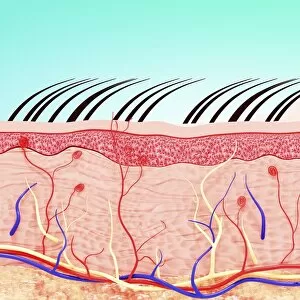
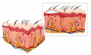
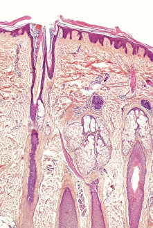
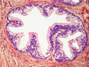
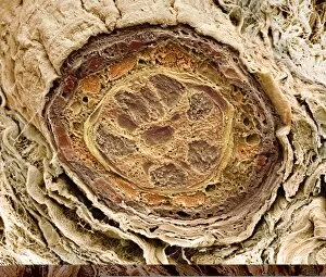
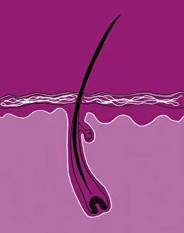
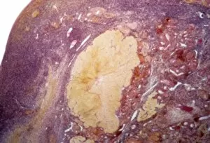
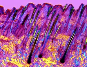
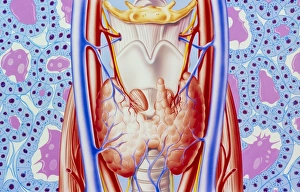
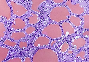
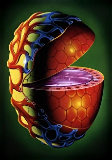
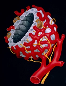
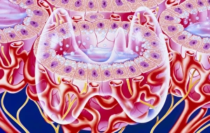
"Follicle: The Hidden World Within Our Skin" Delving beneath the surface, a skin cross section reveals an unwelcome guest - a pesky blackhead lurking within a follicle


All Professionally Made to Order for Quick Shipping
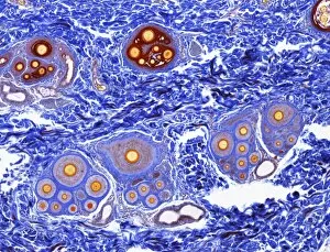
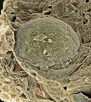
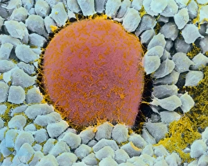
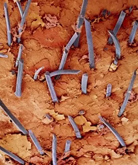
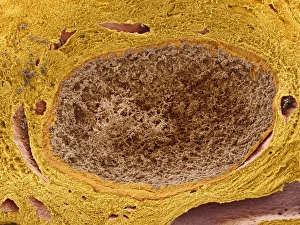
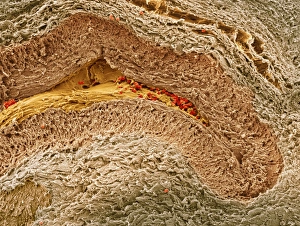
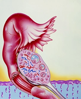
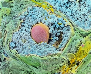
"Follicle: The Hidden World Within Our Skin" Delving beneath the surface, a skin cross section reveals an unwelcome guest - a pesky blackhead lurking within a follicle. A medical illustration showcases the discomforting presence of a pilonidal cyst near the natal cleft of one's buttocks, reminding us of the complexity of our follicles. Diagramming the intricate layers of human skin, we discover that nestled within lies the hair follicle, playing a vital role in our body's protective barrier. Venturing deeper into our anatomy, an illustrated cross-section unveils the fascinating world inside our ovaries - where countless tiny follicles hold potential life. Acne takes various forms; from non-inflammatory to inflammatory types, it reminds us how even these small follicular structures can cause significant distress for some. Conceptual imagery beautifully portrays the layers comprising human skin - with each layer housing its own unique set of functions and intricacies including those related to hair growth. An informative diagram enlightens us about the anatomy of human skin and highlights how crucially intertwined it is with hair follicles in maintaining overall health and protection. Amidst lush landscapes in Kerala, India, Nutmeg trees bear fruit containing precious seeds used since ancient times for their medicinal properties linked to healthy hair follicles. Peering through powerful microscopes reveals another hidden secret - microscopic eyelash mites residing on our lashes' delicate hairs; yet another testament to nature's intricate design within our follicles. Colored SEM images provide mesmerizing glimpses into individual strands adorning our bodies – showcasing both eyelash hairs and sections through hair follicles. Intriguingly complex yet often overlooked, these captions shed light on "follicle, " unraveling its significance across various aspects like skincare concerns or even fertility while inviting appreciation for this hidden world within our skin.