Fraud Blocker Skip to main content

Fluorescent Collection (page 5)

"Unveiling the Vibrant World of Fluorescent Cell Imaging: From Cerebellum Tissue to Dividing Cells" Illuminating the intricate beauty within our brains

Background imageFluorescent Collection: Mexico, Guerrero, Zihuatanejo. Tourist Market- Beach Toys

Mexico, Guerrero, Zihuatanejo. Tourist Market- Beach Toys

Background imageFluorescent Collection: Tokyo, Japan. Tokyo Tower is a communications and observation tower located in Shiba Park

Tokyo, Japan. Tokyo Tower is a communications and observation tower located in Shiba Park, Minato, Tokyo, Japan. At 332.5 metres (1, 091 ft), it is the second tallest artificial structure in Japan

Background imageFluorescent Collection: Indonesia. Fluorsecense in coral (Goniopora sp. )

Indonesia. Fluorsecense in coral (Goniopora sp. )
Indonesia. Fluorsecense in coral (Goniopora sp.)

Background imageFluorescent Collection: Indonesia. Fluorescence in Tube Anemone (Ceranth idai)

Indonesia. Fluorescence in Tube Anemone (Ceranth idai)

Background imageFluorescent Collection: Natural occuring Fluorescence emitted and captured using special barrier filters

Natural occuring Fluorescence emitted and captured using special barrier filters over powerful underwater strobes. Indonesia, Raja Ampat region of Papua (formerly Irian Jaya)

Background imageFluorescent Collection: Metropolitan Police communications room, London

Metropolitan Police communications room, London
View of a Metropolitan Police communications room, showing three officers sitting at desks on the right. There are several push button telephones

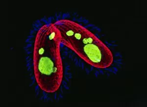
Background imageFluorescent Collection: Tetrahymena protozoa

Tetrahymena protozoa. Immunofluorescent light micrograph of two Tetrahymena thermophila protozoa (single-celled animals). Nuclei are green, cell walls red and cilia (hairs) blue. T

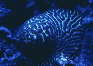
Background imageFluorescent Collection: Fluorescing coral

Fluorescing coral

Background imageFluorescent Collection: Mitosis, fluorescence micrograph

Mitosis, fluorescence micrograph
Mitosis. Fluorescence micrograph of a cell (centre) in anaphase during mitosis (nuclear division). During mitosis two daughter nuclei are formed from one parent nucleus

Background imageFluorescent Collection: Embryonic stem cells

Embryonic stem cells. Computer-enhanced confocal light micrograph of dividing stem cells from the ventricular zone of the retina of a developing embryo

Background imageFluorescent Collection: Abnormal mitosis

Abnormal mitosis
Mitosis. Fluorescence micrograph of a cell during abnormal anaphase of mitosis (nuclear division). During mitosis two daughter nuclei are formed from one parent nucleus

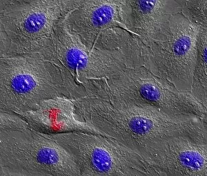
Background imageFluorescent Collection: Dividing cell

Dividing cell. Differential interference contrast (DIC) light micrograph of a cell (lower left) in the metaphase stage of mitosis (cell division). The cells nuclei are stained with fluorescent dye

Background imageFluorescent Collection: Dividing cell, light micrograph

Dividing cell, light micrograph
Cell division. Fluorescent light micrograph of a cell that has divided by mitosis, the asexual replication of a cell into two new cells

Background imageFluorescent Collection: Mitosis

Mitosis. Fluorescence micrograph of a cell during prometaphase of mitosis (nuclear division). During mitosis two daughter nuclei are formed from one parent nucleus

Background imageFluorescent Collection: Fibroblast cells, light micrograph

Fibroblast cells, light micrograph
Fibroblast cells. Confocal light micrograph of human fibroblast cells. The cell nuclei, which contain the cells genetic information, are red

Background imageFluorescent Collection: Brunners glands in the duodenum

Brunners glands in the duodenum
Brunners glands. Fluorescence confocal light micrograph of a section through a human duodenum (small intestine) showing Brunners glands (blue)

Background imageFluorescent Collection: Small intestine

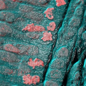
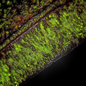
Small intestine. Fluorescence confocal light micrograph of a horizontal section through the mucosa of the human small intestine, showing crypts of Lieberkuhn (pink and blue)

Background imageFluorescent Collection: Colon lining

Colon lining. Fluorescence confocal light micrograph of the lining of a mouse colon (large intestine). The colon starts at the small intestine and ends at the rectum

Background imageFluorescent Collection: White blood cell response

White blood cell response
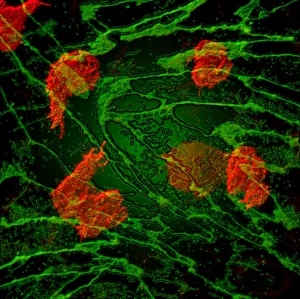
White blood cell movement. Computer-enhanced confocal light micrograph of white blood cells (red) moving through the intact walls of a blood vessel, a process known as diapedesis

Background imageFluorescent Collection: Edison screw fitting

Edison screw fitting of an energy-saving light bulb. This is a type of light bulb connector developed by Thomas Edison in 1909

Background imageFluorescent Collection: Bacterial contamination test

Bacterial contamination test. Ultraviolet (UV) light being used to reveal bacterial contamination. At right, a laboratory workers hand has been placed under a UV light source (pink)

Background imageFluorescent Collection: Banknote security

Banknote security. Euro banknote in ultraviolet light. This highlights security features intended to prevent fraud. Fluorescent ink and strips glow bright blue and red under ultraviolet light

Background imageFluorescent Collection: Energy saving light bulbs

Energy saving light bulbs in the sky, composite image

Background imageFluorescent Collection: Inner ear sensory hairs

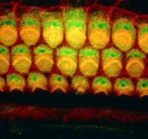
Inner ear sensory hairs
Inner ear hair cells. Confocal light micrograph of V-shaped rows of hair cells (bright arcs) in the organ of Corti. This structure lies in the cochlea of the inner ear

Background imageFluorescent Collection: Purkinje nerve cell, light micrograph

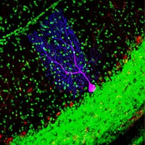
Purkinje nerve cell, light micrograph
Purkinje nerve cell. Confocal light micrograph of a purkinje cell (stained with green fluorescent protein), a type of neuron (nerve cell)

Background imageFluorescent Collection: Nerve cell growth

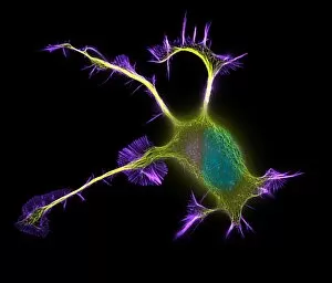
Nerve cell growth. Fluorescent light micrograph of a PC12 cell following stimulation by nerve growth factor. The cell body contains the nucleus (green)

Background imageFluorescent Collection: Purkinje nerve cell

Purkinje nerve cell

Background imageFluorescent Collection: Muscle fibres

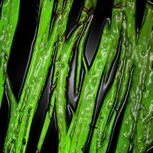
Muscle fibres. Computer-enhanced confocal light micrograph of skeletal (striated) muscle fibres. Skeletal muscle is under the conscious control of the brain

Background imageFluorescent Collection: Looking for biological stains

Looking for biological stains. Forensic scientist using an ultraviolet (UV) lamp to look for stains of biological origin, such as blood, sweat, semen or fingerprints

Background imageFluorescent Collection: Fluorescent dye penetrant test results

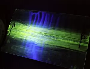
Fluorescent dye penetrant test results
Fluorescent dye penetrant test on a metallic component, viewed under ultraviolet (UV) light. This is a form of non-destructive testing in which a dye is smeared onto the component

Background imageFluorescent Collection: Electricity flowing, artwork

Electricity flowing, artwork
Computer artwork depicting electricity flowing in a fluorescent liquid or gaz or other medium

Background imageFluorescent Collection: Programmed cell death

Programmed cell death. Comparison of a fluorescent light micrograph (top) and transmission electron micrograph (TEM, bottom) of programmed cell death (apoptosis)

Background imageFluorescent Collection: Cultured cell, light micrograph

Cultured cell, light micrograph
Cultured cell. Immunofluorescent light micrograph of an epithelial cell (round, upper right). The cell proteins are marked by fluorescent dyes

Background imageFluorescent Collection: HaCaT culture cells

HaCaT culture cells
HaCaT cells. Immunofluorescence light micrograph of three HaCaT cells. Their nuclei, which contain the cells genetic information in the form of deoxyribonucleic acid (DNA), are blue

Background imageFluorescent Collection: Cell death

Cell death. Computer-enhanced confocal light micrograph of cells in the retina of the eye undergoing programmed cell death (apoptosis)


All Professionally Made to Order for Quick Shipping

"Unveiling the Vibrant World of Fluorescent Cell Imaging: From Cerebellum Tissue to Dividing Cells" Illuminating the intricate beauty within our brains, a light micrograph reveals the mesmerizing patterns of cerebellum tissue. Dive into the microscopic realm as glial cells come alive under a confocal light micrograph, showcasing their fluorescent brilliance. HeLa cells take center stage in this captivating light micrograph (C017 / 8299), displaying their striking fluorescence and cellular structure. Witness another stunning view of HeLa cells (C017 / 8298) through a microscope lens, where their vibrant hues reveal hidden secrets within. Journey into the cerebral cortex and marvel at its nerve cells' fluorescent glow, illuminating the complexity of our brain's architecture. Witness the miracle of life itself as cell division unfolds before your eyes in this breathtaking fluorescent micrograph. Returning to explore cerebellum tissue once more, a light micrograph captures its ethereal essence with radiant colors dancing across each frame. The Metropolitan Police launch on the River Thames may seem unrelated but shares an unexpected connection - fluorescence can be found everywhere. Behold nature's perpetual cycle as dividing cells showcase their vivid fluorescence in yet another awe-inspiring image captured by microscopy techniques. Intriguing and visually stunning, these fluorescent hints offer glimpses into various aspects of cellular biology and beyond – from delicate tissues to bustling cityscapes – reminding us that even at microscopic levels, there is beauty all around us waiting to be discovered.