Fraud Blocker Skip to main content

Fluorescent Collection (page 4)

"Unveiling the Vibrant World of Fluorescent Cell Imaging: From Cerebellum Tissue to Dividing Cells" Illuminating the intricate beauty within our brains

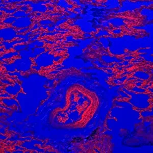
Background imageFluorescent Collection: Lung tissue, confocal micrograph C014 / 4616

Lung tissue, confocal micrograph C014 / 4616
Lung tissue. Laser scanning confocal micrograph (LSCM) of a section through a human lung

Background imageFluorescent Collection: Kidney tissue, confocal micrograph C014 / 4611

Kidney tissue, confocal micrograph C014 / 4611
Kidney tissue. Laser scanning confocal micrograph (LSCM) of a section through a human kidney, showing the cell nuclei (dark)

Background imageFluorescent Collection: Kidney blood vessels, confocal micrograph C014 / 4610

Kidney blood vessels, confocal micrograph C014 / 4610
Kidney blood vessels. Laser scanning confocal micrograph (LSCM) of a section through a human kidney, showing a blood vessel (vertical, centre)

Background imageFluorescent Collection: Arabidopsis thaliana embryo, micrograph C014 / 4590

Arabidopsis thaliana embryo, micrograph C014 / 4590
Arabidopsis thaliana embryo. Confocal light micrograph of a section through the embryo of a thale cress (Arabidopsis thaliana) plant

Background imageFluorescent Collection: Fluorescent imaging fluids C015 / 6515

Fluorescent imaging fluids C015 / 6515
Fluorescent imaging fluids. Lab worker holding flasks of fluorescent fluids used in fluorescent imaging for surgical procedures

Background imageFluorescent Collection: Fluorescent imaging fluids C015 / 6516

Fluorescent imaging fluids C015 / 6516
Fluorescent imaging fluids. Lab worker holding a vial containing a fluorescent fluid used in fluorescent imaging for surgical procedures

Background imageFluorescent Collection: Children receiving light therapy C015 / 6205

Children receiving light therapy C015 / 6205
Phototherapy. A group of children receiving light therapy (phototherapy). A combination of fluorescent and incandescent lamps are being used

Background imageFluorescent Collection: Municipal worker clearing snow C018 / 2338

Municipal worker clearing snow C018 / 2338
Winter. A municipal worker clearing snow from a courtyard. Photographed in Moscow, Russia

Background imageFluorescent Collection: Thale cress anther and pollen

Thale cress anther and pollen. Confocal laser scanning micrograph of pollen grains (round) on a thale cress (Arabidopsis thaliana) anther
Background imageFluorescent Collection: Fibroblast cell, fluorescent micrograph

Fibroblast cell, fluorescent micrograph
Fibroblast cell. Confocal laser scanning micrograph of a fibroblast cell. The cell nucleus, which contains the cells genetic information, is green

Background imageFluorescent Collection: Plant protoplast, fluorescent micrograph

Plant protoplast, fluorescent micrograph
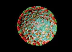
Protoplast. Confocal laser scanning micrograph of a tobacco plant (Nicotiana tabacum) protoplast. A protoplast is a plant cell that has had its tough outer cell wall removed by chemical treatment

Background imageFluorescent Collection: HeLa cells, light micrograph C013 / 4774

HeLa cells, light micrograph C013 / 4774
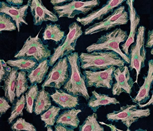
HeLa cells. Multi-photon fluorescence light micrograph of a group of cultured HeLa cells, showing the cell nuclei, which contain the cells genetic information (DNA, red)

Background imageFluorescent Collection: HeLa cells, light micrograph C013 / 4773

HeLa cells, light micrograph C013 / 4773
HeLa cells. Multi-photon fluorescence light micrograph of a group of cultured HeLa cells, showing the cell nuclei, which contain the cells genetic information (DNA, blue), and microtubules (pink)

Background imageFluorescent Collection: Natural ruby crystal

Natural ruby crystal
This ruby is a staggering 1, 085 carats. Mined in Burmas Mogok (Myanmar s) mines and bought by the Museum in 1924 from Burma Ruby Mines Ltd. It is one of the largest crystals in the Museum collection

Background imageFluorescent Collection: Susannite on Caledonite

Susannite on Caledonite
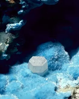
4mm crystal of susannite (carbonate) in a mass of caledonite (suplhate). Specimen from the Roughton Gill mine, Caldbeck Fells, Cumbria

Background imageFluorescent Collection: Fluorite, pale green octahedron

Fluorite, pale green octahedron
With calcite, galena and pyrite. Its properties include fluorescence and cubed crystals. Specimen from Glengowla East mine, Oughterard, Co. Galway, Eire

Background imageFluorescent Collection: Scheelite

Scheelite
Crystals of scheelite (calcium tungstate), an important ore of tungsten from the collections of the Natural History Museum, London

Background imageFluorescent Collection: Diamonds fluorescing

Diamonds fluorescing
Murchison snuff box set with diamonds fluorescing under ultra violet radiation. Gold snuff box set with diamonds ranging from 0.75 to 2.5 carats, presented to Sir R. I. Murchison by Tsar Alexander II

Background imageFluorescent Collection: Celestite

Celestite
A mass of celestite crystals. Celestite is strontium sulphate and is the source of red colours in fireworks

Background imageFluorescent Collection: Autunite

Autunite comprises of (hydrated calcium uranyl phosphate). This is a green, radioactive, highly fluorescent mineral. This specimen is from the Natural History Museum, London

Background imageFluorescent Collection: Franklinite, fluorescing

Franklinite, fluorescing
Ore of zinc fluorescing under ultra violet radiation: calcite - pink; willemite - green; zincite - blue; franklinite - black. See image number 388 for white light view

Background imageFluorescent Collection: Willemite

Willemite
A specimen of willemite (zinc silicate) photographed under UV light

Background imageFluorescent Collection: Torbernite

Torbernite

Background imageFluorescent Collection: Opal doublets

Opal doublets
Opals are not truely crystalline and are therefore mineraloids. They comprise of (hydrated silica glass)

Background imageFluorescent Collection: Adamite

Adamite
A specimen of the mineral adamite (zinc arsenate hydroxide). It is a fluorescent mineral, popular among collectors and shows bright green fluorescent under UV lights

Background imageFluorescent Collection: Fluorescent group

Fluorescent group
A group including amber, ruby, ivory, fluorite and opal photographed in ultra violet light. See 179 for the same group in white light

Background imageFluorescent Collection: Energy efficient light bulbs or Saverlamp, Compact fluorescent energy efficient spiral light bulbs

Energy efficient light bulbs or Saverlamp, Compact fluorescent energy efficient spiral light bulbs or Saverlamps use
MAB-87 Energy efficient light bulbs or Saverlamp Compact fluorescent energy efficient spiral light bulbs or Saverlamps use 80% less energy than traditional bulbs and last up to 10 times longer

Background imageFluorescent Collection: Hundreds of used light bulbs and low energy lamps in recycling bin Cory Environmental landfill

Hundreds of used light bulbs and low energy lamps in recycling bin Cory Environmental landfill site Gloucestershire UK
MAB-652 Hundreds of used light bulbs and low energy lamps in recycling bin Cory Environmental landfill site Gloucestershire UK Mark Boulton contact details: prints@ardea.com tel: +44 (0) 20 8318 1401

Background imageFluorescent Collection: Mexico, Guerrero, Zihuatanejo. Tourist Market- Beach Toys

Mexico, Guerrero, Zihuatanejo. Tourist Market- Beach Toys

Background imageFluorescent Collection: Tokyo, Japan. Tokyo Tower is a communications and observation tower located in Shiba Park

Tokyo, Japan. Tokyo Tower is a communications and observation tower located in Shiba Park, Minato, Tokyo, Japan. At 332.5 metres (1, 091 ft), it is the second tallest artificial structure in Japan

Background imageFluorescent Collection: Indonesia. Fluorsecense in coral (Goniopora sp. )

Indonesia. Fluorsecense in coral (Goniopora sp. )
Indonesia. Fluorsecense in coral (Goniopora sp.)

Background imageFluorescent Collection: Indonesia. Fluorescence in Tube Anemone (Ceranth idai)

Indonesia. Fluorescence in Tube Anemone (Ceranth idai)

Background imageFluorescent Collection: Natural occuring Fluorescence emitted and captured using special barrier filters

Natural occuring Fluorescence emitted and captured using special barrier filters over powerful underwater strobes. Indonesia, Raja Ampat region of Papua (formerly Irian Jaya)

Background imageFluorescent Collection: Metropolitan Police communications room, London

Metropolitan Police communications room, London
View of a Metropolitan Police communications room, showing three officers sitting at desks on the right. There are several push button telephones

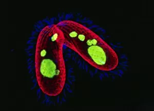
Background imageFluorescent Collection: Tetrahymena protozoa

Tetrahymena protozoa. Immunofluorescent light micrograph of two Tetrahymena thermophila protozoa (single-celled animals). Nuclei are green, cell walls red and cilia (hairs) blue. T

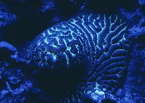
Background imageFluorescent Collection: Fluorescing coral

Fluorescing coral

Background imageFluorescent Collection: Mitosis, fluorescence micrograph

Mitosis, fluorescence micrograph
Mitosis. Fluorescence micrograph of a cell (centre) in anaphase during mitosis (nuclear division). During mitosis two daughter nuclei are formed from one parent nucleus

Background imageFluorescent Collection: Embryonic stem cells

Embryonic stem cells. Computer-enhanced confocal light micrograph of dividing stem cells from the ventricular zone of the retina of a developing embryo

Background imageFluorescent Collection: Abnormal mitosis

Abnormal mitosis
Mitosis. Fluorescence micrograph of a cell during abnormal anaphase of mitosis (nuclear division). During mitosis two daughter nuclei are formed from one parent nucleus

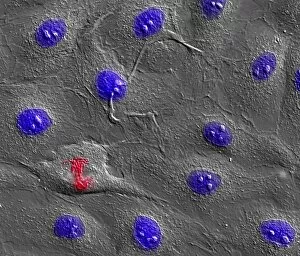
Background imageFluorescent Collection: Dividing cell

Dividing cell. Differential interference contrast (DIC) light micrograph of a cell (lower left) in the metaphase stage of mitosis (cell division). The cells nuclei are stained with fluorescent dye

Background imageFluorescent Collection: Dividing cell, light micrograph

Dividing cell, light micrograph
Cell division. Fluorescent light micrograph of a cell that has divided by mitosis, the asexual replication of a cell into two new cells

Background imageFluorescent Collection: Mitosis

Mitosis. Fluorescence micrograph of a cell during prometaphase of mitosis (nuclear division). During mitosis two daughter nuclei are formed from one parent nucleus

Background imageFluorescent Collection: Fibroblast cells, light micrograph

Fibroblast cells, light micrograph
Fibroblast cells. Confocal light micrograph of human fibroblast cells. The cell nuclei, which contain the cells genetic information, are red

Background imageFluorescent Collection: Brunners glands in the duodenum

Brunners glands in the duodenum
Brunners glands. Fluorescence confocal light micrograph of a section through a human duodenum (small intestine) showing Brunners glands (blue)

Background imageFluorescent Collection: Small intestine

Small intestine. Fluorescence confocal light micrograph of a horizontal section through the mucosa of the human small intestine, showing crypts of Lieberkuhn (pink and blue)

Background imageFluorescent Collection: Colon lining

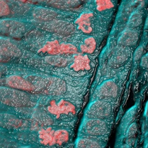
Colon lining. Fluorescence confocal light micrograph of the lining of a mouse colon (large intestine). The colon starts at the small intestine and ends at the rectum

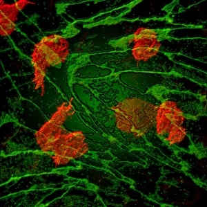
Background imageFluorescent Collection: White blood cell response

White blood cell response
White blood cell movement. Computer-enhanced confocal light micrograph of white blood cells (red) moving through the intact walls of a blood vessel, a process known as diapedesis


All Professionally Made to Order for Quick Shipping

"Unveiling the Vibrant World of Fluorescent Cell Imaging: From Cerebellum Tissue to Dividing Cells" Illuminating the intricate beauty within our brains, a light micrograph reveals the mesmerizing patterns of cerebellum tissue. Dive into the microscopic realm as glial cells come alive under a confocal light micrograph, showcasing their fluorescent brilliance. HeLa cells take center stage in this captivating light micrograph (C017 / 8299), displaying their striking fluorescence and cellular structure. Witness another stunning view of HeLa cells (C017 / 8298) through a microscope lens, where their vibrant hues reveal hidden secrets within. Journey into the cerebral cortex and marvel at its nerve cells' fluorescent glow, illuminating the complexity of our brain's architecture. Witness the miracle of life itself as cell division unfolds before your eyes in this breathtaking fluorescent micrograph. Returning to explore cerebellum tissue once more, a light micrograph captures its ethereal essence with radiant colors dancing across each frame. The Metropolitan Police launch on the River Thames may seem unrelated but shares an unexpected connection - fluorescence can be found everywhere. Behold nature's perpetual cycle as dividing cells showcase their vivid fluorescence in yet another awe-inspiring image captured by microscopy techniques. Intriguing and visually stunning, these fluorescent hints offer glimpses into various aspects of cellular biology and beyond – from delicate tissues to bustling cityscapes – reminding us that even at microscopic levels, there is beauty all around us waiting to be discovered.