
Digestive Tract Collection
The digestive tract, also known as the gastrointestinal tract, is a complex system found in various organisms


All Professionally Made to Order for Quick Shipping
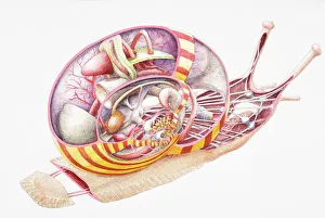
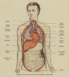
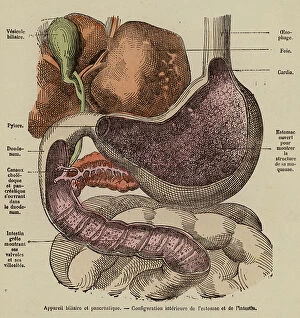
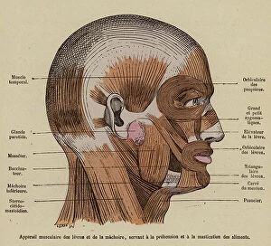
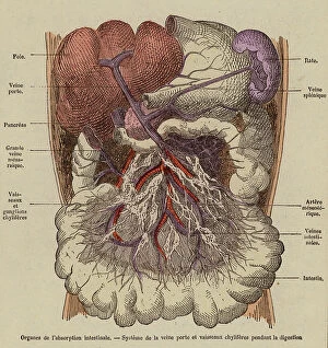
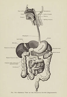
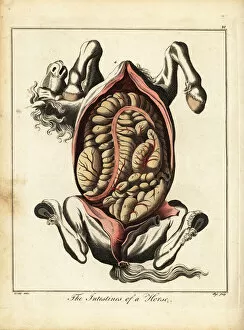
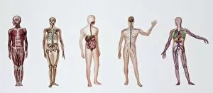
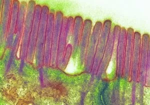
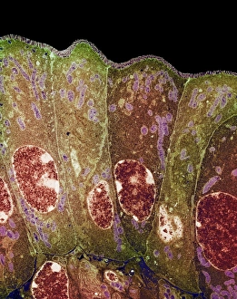
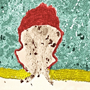
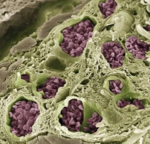
The digestive tract, also known as the gastrointestinal tract, is a complex system found in various organisms. From land snails to horses and even humans, this internal anatomy plays a crucial role in the process of digestion. Imagine taking a cross-section view of a land snail's digestive tract. You would witness an intricate network of organs working together to break down food particles and extract nutrients. Similarly, an engraving from 1792 showcases the intestines of a horse, highlighting their importance in nutrient absorption. In "A doctor's Sunday rest, " we catch a glimpse of how physicians appreciate the significance of maintaining a healthy digestive system for overall well-being. The image portrays their understanding that proper functioning of this vital system is essential for good health. Looking at another perspective, we see a detailed view illustrating the entire digestive tract. This visual representation emphasizes its complexity and interconnectedness with other bodily systems. Biomedical illustrations provide further insight into specific processes within the digestive tract. One such illustration depicts enzymes separating from molecules and passing through the wall into the bloodstream—a fascinating mechanism responsible for nutrient absorption. Another biomedical illustration shows how these powerful enzymes combine with large food molecules like proteins, breaking them down into smaller components that can be easily absorbed by our bodies. Conceptual images highlight different aspects of this remarkable system—such as simple columnar epithelium—an important tissue lining our intestines that aids in nutrient absorption while providing protection against harmful substances. These visuals also remind us that our internal systems are intricately connected; they work together harmoniously to ensure optimal health. Illustrations showcasing various internal systems emphasize this interdependence between different parts of our body. Lastly, artwork depicting healthy large intestines serves as inspiration for maintaining good gut health—a crucial aspect often overlooked but critical for overall well-being. These vibrant depictions encourage us to prioritize caring for our digestive tracts through balanced diets and lifestyle choices. Exploring these captivating visuals allows us to appreciate the complexity and significance of the digestive tract.