Fraud Blocker Skip to main content

Anatomy Collection (page 4)

"Unveiling the Intricacies of Anatomy: From Sensory Homunculus to Motor Homunculus" Step into the fascinating world of anatomy, where every detail tells a story

Background imageAnatomy Collection: Tarantula spider - close-up of face

Tarantula spider - close-up of face (Trochosa singoriensis). South Tuva, Russia

Background imageAnatomy Collection: Neanderthal skull

Neanderthal skull
Skull of an adult, female neanderthal

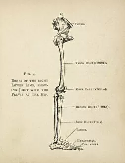
Background imageAnatomy Collection: Diagram of the bones of the right leg and hip

Diagram of the bones of the right leg and hip
Diagram of the bones of the right leg, showing the joint with the pelvis at the hip

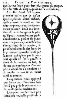
Background imageAnatomy Collection: Homunculus depicted inside human sperm. Woodcut from the 17th century Dutch microsopist

Homunculus depicted inside human sperm. Woodcut from the 17th century Dutch microsopist
HUMAN SPERM, 17th CENTURY. Homunculus depicted inside human sperm. Woodcut from the 17th century Dutch microsopist, Niklass Hartsoekers treatise on microscopy, Essay de Dioptrique

Background imageAnatomy Collection: Da Vinci Quote Simplicity

Da Vinci Quote Simplicity
Fadil Roze

Background imageAnatomy Collection: The Sacred Heart of Jesus, 19th Century (colour litho)

The Sacred Heart of Jesus, 19th Century (colour litho)
942919 The Sacred Heart of Jesus, 19th Century (colour litho) by French School, (19th century); Private Collection; (add.info)

Background imageAnatomy Collection: Three Studies of Horses, c1480 (1945). Artist: Leonardo da Vinci

Three Studies of Horses, c1480 (1945). Artist: Leonardo da Vinci
Three Studies of Horses, c1480 (1945). From The Drawings of Leonardo da Vinci. [Reynal & Hitchcock, New York, 1945]

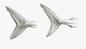
Background imageAnatomy Collection: Black and white illustration of two shark tail fins, single-keeled tail of Mako shark (Isurus sp)

Black and white illustration of two shark tail fins, single-keeled tail of Mako shark (Isurus sp)
Black and white illustration of two shark tail fins, single-keeled tail of Mako shark (Isurus sp.), and double-keeled tail of Porbeagle shark (Lamna nasus)

Background imageAnatomy Collection: Respiratory system, cross-section, drawing

Respiratory system, cross-section, drawing
Medicine - Anatomy - Respiratory system. Drawing, section

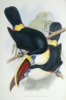
Background imageAnatomy Collection: Ramphastos vitillenus, channel-billed toucan

Ramphastos vitillenus, channel-billed toucan

Background imageAnatomy Collection: Shark Teeth & Jaws

Shark Teeth & Jaws
PM-9069 Shark Teeth & Jaws Odontaspis Pat Morris Please note that prints are for personal display purposes only and may not be reproduced in any way

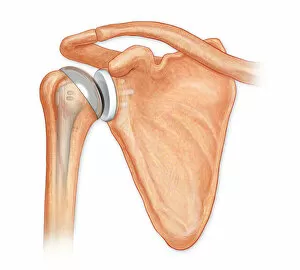
Background imageAnatomy Collection: Anterior view total shoulder joint repair

Anterior view total shoulder joint repair
Horizontal, Detail, Anatomy, Diagram, Repair, Illustration, Bone, Shoulder, Biology, Joint, Cut Out, White Background, Artwork, Color Image, Front View, Close Up, Healthcare And Medicine

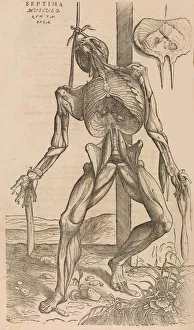
Background imageAnatomy Collection: De humani corporis fabrica (Of the Structure of the Human Body), 1555

De humani corporis fabrica (Of the Structure of the Human Body), 1555

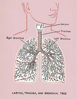
Background imageAnatomy Collection: Lung Diagram

Lung Diagram
http://csaimages.com/images/istockprofile/csa_vector_dsp.jpg

Background imageAnatomy Collection: Achilles tendon

Achilles tendon
achilles tendon, anatomy, back view, calcaneal tendon, human, illustr, Medical Image Collection, 87320008

Background imageAnatomy Collection: Anatomy of Organs Engraving

Anatomy of Organs Engraving
Engraved illustrations of Anatomy of the Fasciae, Integuents, and Organs of Mastication and Respiration from Iconographic Encyclopedia of Science, Literature and Art, Published in 1851

Background imageAnatomy Collection: Study of a Rearing Horse, c1480 (1945). Artist: Leonardo da Vinci

Study of a Rearing Horse, c1480 (1945). Artist: Leonardo da Vinci
Study of a Rearing Horse, c1480 (1945). From The Drawings of Leonardo da Vinci. [Reynal & Hitchcock, New York, 1945]

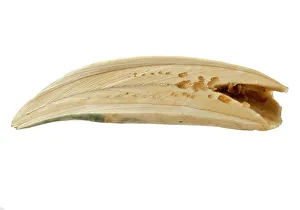
Background imageAnatomy Collection: Physeter macrocephalus, Sperm whale tooth

Physeter macrocephalus, Sperm whale tooth
Sperm whale tooth

Background imageAnatomy Collection: Urania speciosa; Palm

Urania speciosa; Palm
Watercolour from the John Reeves Collection; c.1820s Date: 1820

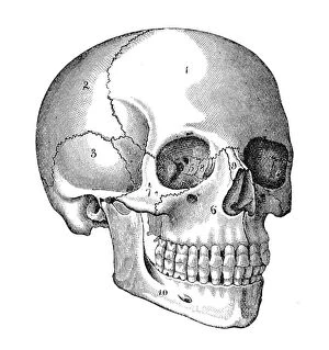
Background imageAnatomy Collection: Human Skull

Human Skull
Illustration of human skull

Background imageAnatomy Collection: Maxilla bone with teeth

Maxilla bone with teeth
Illustration engraving of a maxilla bone with teeth

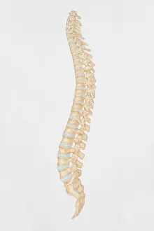
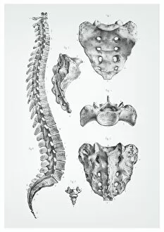
Background imageAnatomy Collection: Diagram of the human spine, side view

Diagram of the human spine, side view

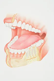
Background imageAnatomy Collection: Cross-section diagram of human mouth and jaw

Cross-section diagram of human mouth and jaw

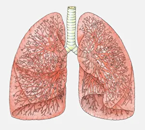
Background imageAnatomy Collection: Diagram of human lungs showing blood supply

Diagram of human lungs showing blood supply

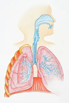
Background imageAnatomy Collection: Illustration of human respiratory system showing oral cavity, and nasal cavity, larynx, trachea

Illustration of human respiratory system showing oral cavity, and nasal cavity, larynx, trachea, bronchus, and lungs

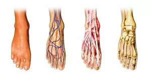
Background imageAnatomy Collection: Human foot anatomy showing skin, veins, arteries, muscles and bones

Human foot anatomy showing skin, veins, arteries, muscles and bones
Human foot anatomy showing skin, veins, arteries, muscles, and bones, cutaway view

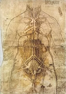
Background imageAnatomy Collection: Leonardo Da Vinci: Anatomy

Leonardo Da Vinci: Anatomy
LEONARDO DA VINCI: ANATOMY. Study of female anatomy

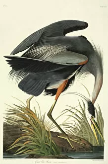
Background imageAnatomy Collection: Ardea herodias, great blue heron

Ardea herodias, great blue heron
Plate 211 from John James Audubons Birds of America, original double elephant folio (1834-35), hand-coloured aquatint. Engraved, printed and coloured by R. Havell (& Son), London

Background imageAnatomy Collection: EUSTACHIO, Bartolomeo (1500-1574). Italian anatomist

EUSTACHIO, Bartolomeo (1500-1574). Italian anatomist

Background imageAnatomy Collection: Araucaria columnaris & A. heterophyllo

Araucaria columnaris & A. heterophyllo - Coloured copper engraving by Warner based on watercolours by Ferdinand Bauer, A.B. Lambert, A description of the genus Pinus, London, 1803, t.39 Date: 1803

Background imageAnatomy Collection: Bungarus fasciatus, Banded Krait

Bungarus fasciatus, Banded Krait
Snake skin mounted on paper, The Patrick Russell Collection c.1790 Date: circa 1790

Background imageAnatomy Collection: Anatomy of the human heart

Anatomy of the human heart.. Anatomy of the human heart. Copperplate engraving by Milton from Abraham Rees Cyclopedia or Universal Dictionary of Arts, Sciences and Literature, Longman, Hurst, Rees

Background imageAnatomy Collection: Human Skull Engraving

Human Skull Engraving

Background imageAnatomy Collection: Human eye anatomy engraving 1899

Human eye anatomy engraving 1899
Corso Elementare di Scienze Naturali

Background imageAnatomy Collection: Human spine anatomy illustration 1866

Human spine anatomy illustration 1866
Atlas d anatomie descriptive du corps humain

Background imageAnatomy Collection: Skeleton of a fowl lithograph 1897

Skeleton of a fowl lithograph 1897
Diseases of Poultry by Leonard Pearson - Clarence M. Busch, State Printer of Pennsylvania 1897
Background imageAnatomy Collection: Hand

Hand
Antique illustration of a human hand with tendons

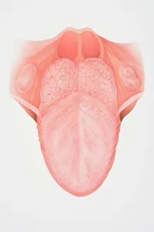
Background imageAnatomy Collection: Diagram illustrating the anatomy of the tongue, front view

Diagram illustrating the anatomy of the tongue, front view

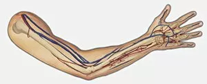
Background imageAnatomy Collection: Diagram showing bones, veins and arteries in a human arm and hand

Diagram showing bones, veins and arteries in a human arm and hand

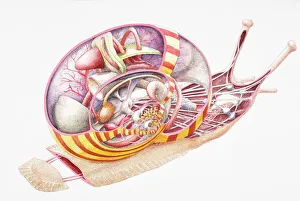
Background imageAnatomy Collection: Land Snail (Gastropoda), internal anatomy, cross-section

Land Snail (Gastropoda), internal anatomy, cross-section

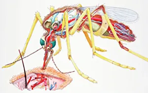
Background imageAnatomy Collection: Mosquito (Culicidae), female, internal anatomy, and sucking blood from skin, cross-section

Mosquito (Culicidae), female, internal anatomy, and sucking blood from skin, cross-section

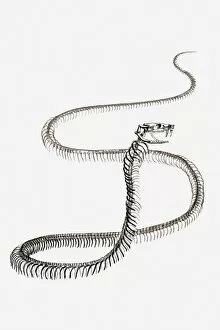
Background imageAnatomy Collection: Black and white illustration of a snakes skeleton

Black and white illustration of a snakes skeleton

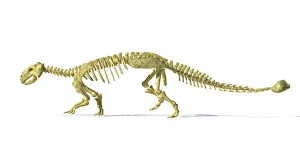
Background imageAnatomy Collection: 3D rendering of an Ankylosaurus dinosaur skeleton

3D rendering of an Ankylosaurus dinosaur skeleton, side view. This armored dinosaur lived in the early Mesozoic era

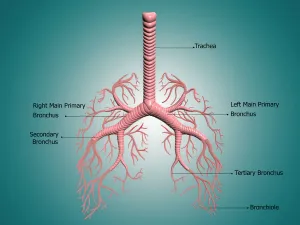
Background imageAnatomy Collection: Anatomy of the bronchus and bronchial tubes

Anatomy of the bronchus and bronchial tubes
Digital illustration showing the structure of the bronchus and bronchial tubes. These pathways carry air into the lungs and carbon dioxide out

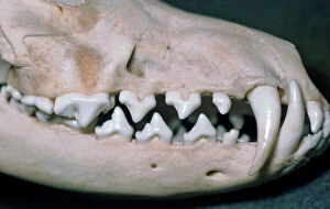
Background imageAnatomy Collection: Picture No. 10864938

Picture No. 10864938
Dog skull showing large canines Date:

Background imageAnatomy Collection: Pen and ink studies by Leonardo da Vinci, c1510-11, of the human hand

Pen and ink studies by Leonardo da Vinci, c1510-11, of the human hand
LEONARDO: HANDS, c1510. Pen and ink studies by Leonardo da Vinci, c1510-11, of the human hand

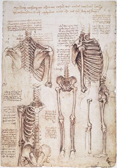
Background imageAnatomy Collection: Pen and ink studies by Leonardo da Vinci, c1510, of human thorax, pelvic, and leg bones

Pen and ink studies by Leonardo da Vinci, c1510, of human thorax, pelvic, and leg bones
LEONARDO: THORAX, c1510. Pen and ink studies by Leonardo da Vinci, c1510, of human thorax, pelvic, and leg bones


All Professionally Made to Order for Quick Shipping

"Unveiling the Intricacies of Anatomy: From Sensory Homunculus to Motor Homunculus" Step into the fascinating world of anatomy, where every detail tells a story. Explore the depths of human and animal structures as we delve into this captivating subject. Let's begin with Leonardo da Vinci's masterpiece, "Head of a Bear. " Created in 1480 but discovered in 1945, this artwork showcases da Vinci's unparalleled ability to capture anatomical accuracy. The intricate details reveal his keen observation skills and deep understanding of form. Moving on to our furry friends, the greyhound takes center stage. Discovering the anatomy of these majestic creatures unveils their remarkable speed and agility. From their sleek skeleton to their muscular build, it is evident why they are renowned for their racing abilities. But what about us humans? Enter the sensory homunculus – a visual representation that depicts how our brain perceives different body parts based on sensitivity levels. This map reveals intriguing insights into how our senses are distributed throughout our bodies. X-ray images provide another dimension to understanding anatomy. Take a glimpse at normal knees through an X-ray lens – marvel at the complexity hidden beneath our skin while appreciating its delicate balance between strength and flexibility. The backbone is often considered one of nature's most ingenious designs. Our human backbone includes ribs and pelvis, providing stability while allowing movement – truly an architectural marvel worth exploring further. Venturing deeper within ourselves, let us explore the intricacies of the human brain from an inferior view. Witness its complex network of connections responsible for controlling various bodily functions - truly awe-inspiring. As we journey back in time, we encounter hominid crania – remnants that shed light on our evolutionary history. These ancient skulls offer glimpses into early forms such as Australopithecus afarensis (AL 288-1), famously known as Lucy - bridging gaps between past and present.