Fraud Blocker Skip to main content

Anatomy Collection (page 5)

"Unveiling the Intricacies of Anatomy: From Sensory Homunculus to Motor Homunculus" Step into the fascinating world of anatomy, where every detail tells a story

Background imageAnatomy Collection: Scolopendra gigantea, giant centipede

Scolopendra gigantea, giant centipede
A giant centipede which was brought into the Museums Insect Identification service after being found in a living room in London. The specimen is venomous and not native to the UK

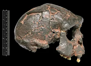
Background imageAnatomy Collection: Homo erectus, Java Man cranium (Sangiran 17) cast

Homo erectus, Java Man cranium (Sangiran 17) cast
Lateral view of partially reconstructed cranium of Homo erectus Java Man about 700, 000 years old known as Sangiran 17. Discovered by Towikromo in 1969

Background imageAnatomy Collection: Riparia riparia, Hirundo rustica, Delichon urbica

Riparia riparia, Hirundo rustica, Delichon urbica
Plate 36 from Archibald Thorburns second edtition of British Birds, Vol. 1 (1925)

Background imageAnatomy Collection: Mammoth skeleton drawing

Mammoth skeleton drawing
Plate 10 from Memoires de L Acadamie Imperiale des Sciences, Vol 5, 1815

Background imageAnatomy Collection: Parasaurolophus skeleton

Parasaurolophus skeleton
This dinosaur which grew up to 10 metres in length had a large crest over the top of its head which extended over a metre. It lived during the Upper Cretaceous period, 76 to 74 mya

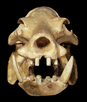
Background imageAnatomy Collection: Skull of a pigmy hippo

Skull of a pigmy hippo
Model of a pigmy hippo skull from Cyprus

Background imageAnatomy Collection: Snake skeleton by Albertus Seba

Snake skeleton by Albertus Seba
Tab 107 illustrating a section of snake skeleton from Thesaurus, by Albertus Seba

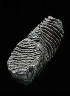
Background imageAnatomy Collection: Tooth from a woolly mammoth

Tooth from a woolly mammoth
Tooth from an extinct woolly mammoth, specimen from the Natural History Museum, London

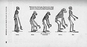
Background imageAnatomy Collection: Hylobates sp. Pongo pygmaeus, Pan troglodytes, Gorilla gori

Hylobates sp. Pongo pygmaeus, Pan troglodytes, Gorilla gori
Gibbon, orangutan, chimpanzee, gorilla and human. Evidence as to Mans Place in Nature. Illustration published in Mans Place in Nature, Vol. 7 from a collection of essays by Thomas Henry Huxley, 1863

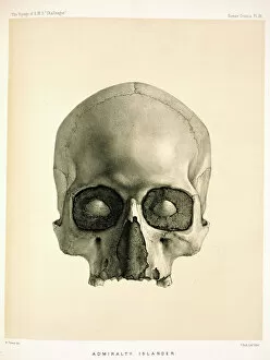
Background imageAnatomy Collection: Engraving of a human skull

Engraving of a human skull
Collected from the Admiralty Islands in the south-western Pacific, on The Voyage of H.M.S. Challenger (1872-1876) was funded by the British Government for the purpose of scientific discovery

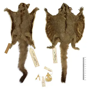
Background imageAnatomy Collection: Petaurus breviceps ariel, sugar glider

Petaurus breviceps ariel, sugar glider
Petaurus breviceps ariel (Gould, 1849) sugar glider. BMNH 1842.5.26.1, female skin & skull, lectotype photographed next to specimen BMNH 1855.12.24.308 paralectotype skin

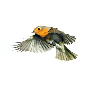
Background imageAnatomy Collection: Erithacus rubecula, European robin

Erithacus rubecula, European robin
An European robin (Erithacus rubecula) in flight, U.K. Photographed by Frank Greenaway

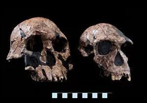
Background imageAnatomy Collection: Homo rudolfensis (KNM-ER 1470) Homo habilis (KNM-ER 1813)

Homo rudolfensis (KNM-ER 1470) Homo habilis (KNM-ER 1813)
On the left, KNM-ER 1470 (also attributed to H. rudolfensis). On the right, KNM-ER 1813. Both skulls are about 2 million years old. Held at The Natural History Museum, London

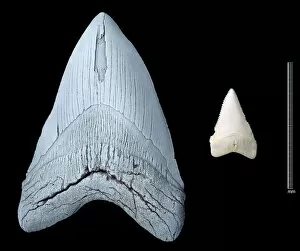
Background imageAnatomy Collection: Sharks teeth

Sharks teeth
Tooth of an extinct shark (Carcharodon megalodon) on the right, compared with a tooth from a modern Great White shark, (Carcharodon carcharias) on the left

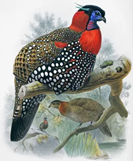
Background imageAnatomy Collection: Tragopan melanocephala, western tragopan

Tragopan melanocephala, western tragopan
Plate 23 by Joseph Wolf from Daniel Giraud Elliots A Monograph of the Phasianid, or Family of the Pheasants, (1872)

Background imageAnatomy Collection: Phasianus colchicus decollatus, common (Chinese ringless) ph

Phasianus colchicus decollatus, common (Chinese ringless) ph
Plate 57 by Joseph Wolf from Daniel Giraud Elliots A Monograph of the Phasianid, or Family of the Pheasants, (1872)

Background imageAnatomy Collection: Turdus philomelos, song thrush

Turdus philomelos, song thrush
Plate 19 from Henry Eeles Dressers A History of the Birds of Europe.. (1871-96)

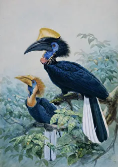
Background imageAnatomy Collection: Ceratagymna elata, yellow-casqued hornbill

Ceratagymna elata, yellow-casqued hornbill
Watercolour and gouache John Gerrard Keulemans

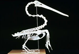
Background imageAnatomy Collection: Brown Pelican Skeleton - coastal California

Brown Pelican Skeleton - coastal California
KEL-1152 Brown Pelican Skeleton - coastal California Distribution includes Trinidad & Tobago and Galapagos Peiecanus occidentalis Ken Lucas Please note that prints are for personal display purposes

Background imageAnatomy Collection: American Alligator Skull - Florida - Southeastern USA

American Alligator Skull - Florida - Southeastern USA
KEL-1189 American Alligator Skull - Florida Southeastern USA Alligator mississippiensis Ken Lucas Please note that prints are for personal display purposes only and may not be reproduced in any way

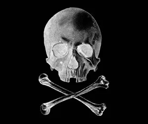
Background imageAnatomy Collection: Skull and Crossbones - Inverted

Skull and Crossbones - Inverted
The Jolly Roger is the name given to any of various flags flown to identify a ships crew as pirates. The flag most usually identified as the Jolly Roger today is the skull and crossbones

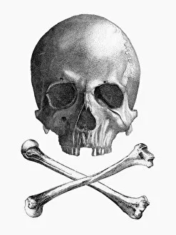
Background imageAnatomy Collection: Skull and Crossbones

Skull and Crossbones
The Jolly Roger is the name given to any of various flags flown to identify a ships crew as pirates. The flag most usually identified as the Jolly Roger today is the skull and crossbones

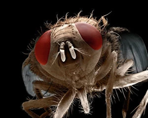
Background imageAnatomy Collection: Scanning Electron Micrograph (SEM): House Fly, Magnification x 40 (A4 size: 29. 7 cm width)

Scanning Electron Micrograph (SEM): House Fly, Magnification x 40 (A4 size: 29. 7 cm width)
LRDS-72 House Fly Scanning Electron Micrograph (SEM) Musca domestica Magnification x 40 (A4 size: 29.7 cm width) Coloured by hand to enhance natural features

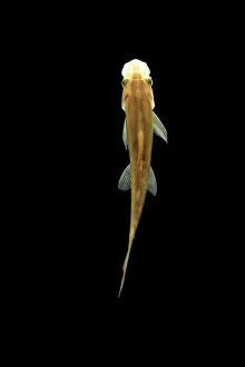
Background imageAnatomy Collection: Fish - Sucking Loach Asia

Fish - Sucking Loach Asia
BB-800 Fish - Sucking Loach / Chinese Algae Eater Distribution: Thailand, Asia Gyrinocheilus aymonieri Brian Bevan Please note that prints are for personal display purposes only

Background imageAnatomy Collection: Broad Bodied Chaser Dragonfly - warming on Yellow Iris Norfolk UK

Broad Bodied Chaser Dragonfly - warming on Yellow Iris Norfolk UK
FEU-234 Broad Bodied Chaser Dragonfly - warming on Yellow Iris Norfolk, UK Libellula depressa Geoff du Feu Please note that prints are for personal display purposes only

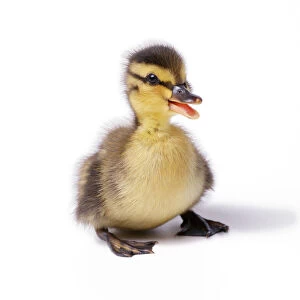
Background imageAnatomy Collection: Mallard Duck - duckling at 24 hours. Sequence 4 of 4

Mallard Duck - duckling at 24 hours. Sequence 4 of 4
JD-15752 Mallard Duck - duckling at 24 hours Sequence 4 of 4 John Daniels Please note that prints are for personal display purposes only and may not be reproduced in any way

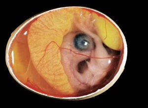
Background imageAnatomy Collection: Chicken chick - 10 day old embryo in egg

Chicken chick - 10 day old embryo in egg
LA-2610 Chicken chick - 10 day old embryo in egg Jean Michel Labat Please note that prints are for personal display purposes only and may not be reproduced in any way

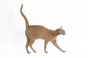
Background imageAnatomy Collection: Fawn Oriental Cat

Fawn Oriental Cat
LA-1993 Fawn Oriental Cat Jean Michel Labat Please note that prints are for personal display purposes only and may not be reproduced in any way. contact details: prints@ardea.com tel: 020 8318 1401

Background imageAnatomy Collection: Heart (Latin)

Heart (Latin)
with its various vessels identified by their latin names

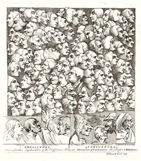
Background imageAnatomy Collection: Hogarth Faces 1807

Hogarth Faces 1807
A myriad of faces looking in different directions: Characters and Caricatures

Background imageAnatomy Collection: Rembrandts Dissection

Rembrandts Dissection
An anatomical demonstration

Background imageAnatomy Collection: Crime / Resurrectionists

Crime / Resurrectionists
Robbing graveyards for corpses which will be sold to medical schools for purposes of anatomy

Background imageAnatomy Collection: Medical College For Women Women Woman Dissecting

Medical College For Women Women Woman Dissecting
A student dissecting a leg at the Medical College for Women on East Twelfth Street and Second Avenue, New York, USA. After an illustration from an 1870 edition of Frank Leslie's Illustrated

Background imageAnatomy Collection: Odin questioning Mimir (colour litho)

Odin questioning Mimir (colour litho)
7173279 Odin questioning Mimir (colour litho) by Doepler, Carl Emil (1824-1905); Private Collection; (add.info.: Odin questioning Mimir)

Background imageAnatomy Collection: Anatomy of the nervous system in the heart, neck and arm

Anatomy of the nervous system in the heart, neck and arm. Lithograph by C. Schach from Lorenz Okens Universal Natural History, Allgemeine Naturgeschichte fur alle Stande, Stuttgart, 1839

Background imageAnatomy Collection: Are you nervous? If so, find the nerve! Photograph of Dr

Are you nervous? If so, find the nerve! Photograph of Dr. Rufus Weaver showing the nervous system of a man. Date: 1906

Background imageAnatomy Collection: The Right Kidney

The Right Kidney
http://csaimages.com/images/istockprofile/csa_vector_dsp.jpg

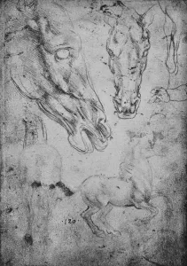
Background imageAnatomy Collection: Studies of Horses and of Horses Heads, c1480 (1945). Artist: Leonardo da Vinci

Studies of Horses and of Horses Heads, c1480 (1945). Artist: Leonardo da Vinci
Studies of Horses and of Horses Heads, c1480 (1945). From The Drawings of Leonardo da Vinci. [Reynal & Hitchcock, New York, 1945]

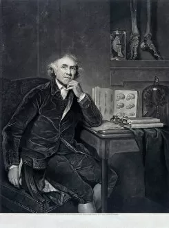
Background imageAnatomy Collection: John Hunter, 1786. Artist: WO Geller

John Hunter, 1786. Artist: WO Geller
John Hunter, 1786. Scottish surgeon and anatomist whose series of lectures on the theory and practice of surgery attracted numerous students, including Edward Jenner, Astley Cooper

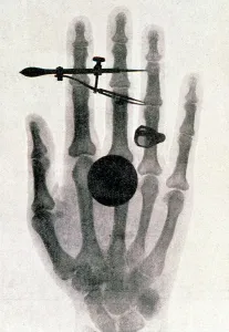
Background imageAnatomy Collection: Wilhelm Roentgens X-ray photograph of his wifes hand, 1896. Artist: Wilhelm Conrad Rontgen

Wilhelm Roentgens X-ray photograph of his wifes hand, 1896. Artist: Wilhelm Conrad Rontgen
Wilhelm Roentgens X-ray photograph of his wifes hand, 1896. For his work on electromagnetic radiation and the discovery of X-rays, Roentgen (1845-1923) was awarded the 1901 Nobel prize for Physics

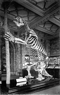
Background imageAnatomy Collection: Giant Ground Sloth, Natural History Museum

Giant Ground Sloth, Natural History Museum
Photograph of a Skeleton of the Giant Ground Sloth (Megatherium Americanum). July 1902. Archive ref: PH/173/244 Date: 1902

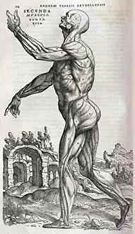
Background imageAnatomy Collection: Anatomical drawing of musculature, side view

Anatomical drawing of musculature, side view
Secunda Musculorum Tabulae (Anatomical drawing of musculature, side view) Source: Andreae Vesalii Bruxellensis, scholae medicorum Patauinae professoris

Background imageAnatomy Collection: Unfolding female anatomy

Unfolding female anatomy
x Date: 19th century

Background imageAnatomy Collection: Repeating Pattern - Skeletons

Repeating Pattern - Skeletons (white background). *PLEASE NOTE that the magnifying glass is solely to show the detail of the repeating pattern and will not feature on the final file.*

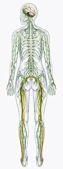
Background imageAnatomy Collection: Digital illustration of human nervous system connected to spinal cord and brain

Digital illustration of human nervous system connected to spinal cord and brain

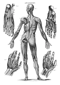
Background imageAnatomy Collection: Human muscles anatomy engraving 1878

Human muscles anatomy engraving 1878
Encyclopedia Britannica 9th Edition Vol I New York, Samuel Hall 1878

Background imageAnatomy Collection: Digital illustration of Bocas area (purple), Wernickes area (orange)

Digital illustration of Bocas area (purple), Wernickes area (orange), Geschwinds territory (turquoise) and arcuate fasciculus (green) in human brain

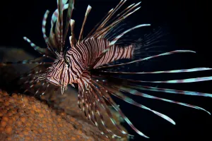
Background imageAnatomy Collection: Red Lionfish flares its deadly spines

Red Lionfish flares its deadly spines as a warning to the photographer not to get any closer, Bonaire, Caribbean Netherlands


All Professionally Made to Order for Quick Shipping

"Unveiling the Intricacies of Anatomy: From Sensory Homunculus to Motor Homunculus" Step into the fascinating world of anatomy, where every detail tells a story. Explore the depths of human and animal structures as we delve into this captivating subject. Let's begin with Leonardo da Vinci's masterpiece, "Head of a Bear. " Created in 1480 but discovered in 1945, this artwork showcases da Vinci's unparalleled ability to capture anatomical accuracy. The intricate details reveal his keen observation skills and deep understanding of form. Moving on to our furry friends, the greyhound takes center stage. Discovering the anatomy of these majestic creatures unveils their remarkable speed and agility. From their sleek skeleton to their muscular build, it is evident why they are renowned for their racing abilities. But what about us humans? Enter the sensory homunculus – a visual representation that depicts how our brain perceives different body parts based on sensitivity levels. This map reveals intriguing insights into how our senses are distributed throughout our bodies. X-ray images provide another dimension to understanding anatomy. Take a glimpse at normal knees through an X-ray lens – marvel at the complexity hidden beneath our skin while appreciating its delicate balance between strength and flexibility. The backbone is often considered one of nature's most ingenious designs. Our human backbone includes ribs and pelvis, providing stability while allowing movement – truly an architectural marvel worth exploring further. Venturing deeper within ourselves, let us explore the intricacies of the human brain from an inferior view. Witness its complex network of connections responsible for controlling various bodily functions - truly awe-inspiring. As we journey back in time, we encounter hominid crania – remnants that shed light on our evolutionary history. These ancient skulls offer glimpses into early forms such as Australopithecus afarensis (AL 288-1), famously known as Lucy - bridging gaps between past and present.