Fraud Blocker Skip to main content

Anatomy Collection (page 3)

"Unveiling the Intricacies of Anatomy: From Sensory Homunculus to Motor Homunculus" Step into the fascinating world of anatomy, where every detail tells a story

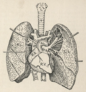
Background imageAnatomy Collection: Diagram of the heart, lungs and windpipe

Diagram of the heart, lungs and windpipe

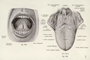
Background imageAnatomy Collection: Anatomy / Head / Tongue

Anatomy / Head / Tongue
MOUTH A medical diagram of the human tongue (lower and upper surfaces), with the specific zones of taste and sensation marked by their Latin names

Background imageAnatomy Collection: Scanning Electron Micrograph (SEM): Fruit Fly, Magnification x 300 (A4 size: 29. 7 cm width)

Scanning Electron Micrograph (SEM): Fruit Fly, Magnification x 300 (A4 size: 29. 7 cm width)
LRDS-57 Fruit Fly Scanning Electron Micrograph (SEM) Drosophila sp Magnification x 300 (A4 size: 29.7 cm width) Coloured by hand to enhance natural features

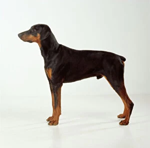
Background imageAnatomy Collection: Dobermann JD 13821 © John Daniels / ARDEA LONDON

Dobermann JD 13821 © John Daniels / ARDEA LONDON
JD-13821 DOG - Dobermann / Doberman Pinscher, standing, side view John Daniels Please note that prints are for personal display purposes only and may not be reproduced in any way

Background imageAnatomy Collection: Maple tree - close-up of leaves & seeds

Maple tree - close-up of leaves & seeds
LA-1525 Sycamore - winged seeds and leaves Studio Jean Michel Labat Please note that prints are for personal display purposes only and may not be reproduced in any way

Background imageAnatomy Collection: Autumn leaf - Underside of leaf

Autumn leaf - Underside of leaf
CLA-149 Autumn leaf - Underside of leaf Colorado Mary Clay Please note that prints are personal display purposes only and may not be reproduced in any way

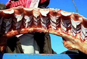
Background imageAnatomy Collection: Jaw of a 4 metre tiger shark, replaceable teeth in 9 rows Egypt Red Sea

Jaw of a 4 metre tiger shark, replaceable teeth in 9 rows Egypt Red Sea
KA-516 Jaw of a 4 metre tiger shark, replaceable teeth in 9 rows Egypt Red Sea Kurt Amsler Please note that prints are for personal display purposes only and may not be reproduced in any way

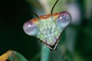
Background imageAnatomy Collection: Praying Mantis - head close-up Kenya, Africa

Praying Mantis - head close-up Kenya, Africa
DOW-88 Praying MANTIS - head close-up Kenya, Africa Pseudocreobotra aeruginosa Steve Downer Please note that prints are for personal display purposes only and may not be reproduced in any way

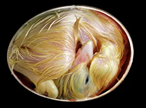
Background imageAnatomy Collection: Chicken chick - 20 day old chick in egg

Chicken chick - 20 day old chick in egg
LA-2611 Chicken chick - 20 day old chick in egg Jean Michel Labat Please note that prints are for personal display purposes only and may not be reproduced in any way

Background imageAnatomy Collection: Cockatiel In flight

Cockatiel In flight
JD-16246 COCKATIEL - in flight Nymphicus hollandicus John Daniels Please note that prints are for personal display purposes only and may not be reproduced in any way

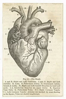
Background imageAnatomy Collection: Heart (Anon)

Heart (Anon)
the heart showing the ventricles, auricles, aorta, arteries, carotid branches and other associated vessels

Background imageAnatomy Collection: Anatomy / Various Parts

Anatomy / Various Parts
Diagrams of various organs, the skull and human body

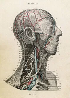
Background imageAnatomy Collection: Anatomy of Head & Throat

Anatomy of Head & Throat
Detailed diagram of the head and throat Date: Nineteenth century

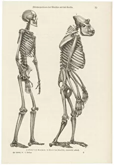
Background imageAnatomy Collection: Two skeletons, human and gorilla

Two skeletons, human and gorilla
A human skeleton compared with that of a gorilla

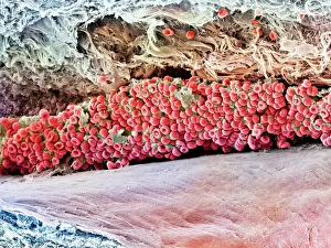
Background imageAnatomy Collection: Scanning electron micrograph (SEM) of red blood cell

Scanning electron micrograph (SEM) of red blood cell
Anatomy, Biology, Blood Clot, Blood Vessel, Cell, Color Image, Fractur, SEM, 85758265

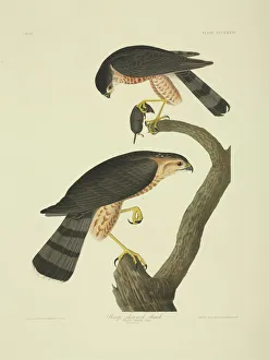
Background imageAnatomy Collection: Accipiter striatus, sharp-shinned hawk

Accipiter striatus, sharp-shinned hawk
Plate 374 from John James Audubons Birds of America, original double elephant folio (1835-38), hand-coloured aquatint. Engraved, printed and coloured by R. Havell (& Son), London

Background imageAnatomy Collection: Skeleton & Ligaments

Skeleton & Ligaments
Physiological diagram of the skeleton and ligaments

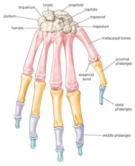
Background imageAnatomy Collection: Bones of the hand

Bones of the hand: the carpal bones (wrist bones), metacarpal bones (bones of the hand proper), and phalanges (finger bones)

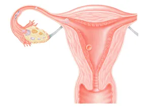
Background imageAnatomy Collection: Digital illustration showing journey of fertilized human egg

Digital illustration showing journey of fertilized human egg

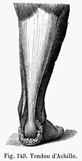
Background imageAnatomy Collection: Line engraving, French, 19th century

Line engraving, French, 19th century
ACHILLES TENDON. Line engraving, French, 19th century

Background imageAnatomy Collection: Troglodytes troglodytes, winter wren

Troglodytes troglodytes, winter wren
Plate 118 from William MacGillivrays Watercolour drawings of British Animals (1831-1841)

Background imageAnatomy Collection: Nyctea scandiaca, snowy owl

Nyctea scandiaca, snowy owl
Plate 34 from John Goulds The Birds of Great Britain, Vol. 1 (1873). Hand coloured lithograph

Background imageAnatomy Collection: Taraxacum officinale, dandelion (fruiting head)

Taraxacum officinale, dandelion (fruiting head)
Scanning electron microscope image showing a vertical section through an unripe fruiting head of a dandelion in the yellow flower stage. Colour added artificially by computer

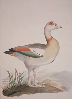
Background imageAnatomy Collection: Alopochen aegyptiaca, Egyptian goose

Alopochen aegyptiaca, Egyptian goose
Plate 5 from William MacGillivrays Watercolour drawings of British Animals (1831-1841)

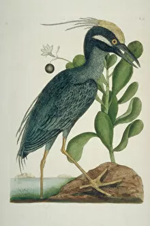
Background imageAnatomy Collection: Nyctanassa violacea, yellow-crowned night heron

Nyctanassa violacea, yellow-crowned night heron
Plate 79, hand coloured etching from The Natural History of Carolina, Florida and the Bahama islands (1731-43) Vol. 1 by Mark Catesby

Background imageAnatomy Collection: Simulium damnosum, Simulian blackfly

Simulium damnosum, Simulian blackfly
Scanning electron microscope image of the head showing the compound eye (x 130). The fly is a vector of a parasite which causes River Blindness. Coloured artifically by computer

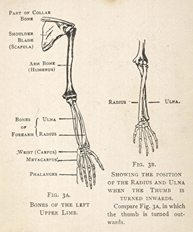
Background imageAnatomy Collection: Diagrams of the bones of hand and arm

Diagrams of the bones of hand and arm
Diagrams of the bones of the left arm and hand, showing the position of the radius and ulna when the thumb is turned inwards. The shoulder blade and part of the collar bone can also be seen

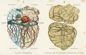
Background imageAnatomy Collection: Heart, Veins, Arteries

Heart, Veins, Arteries
Left - the arteries and veins Right - the muscles

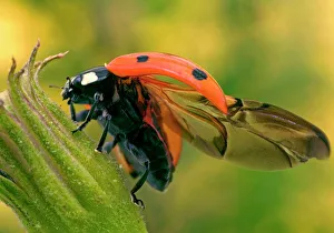
Background imageAnatomy Collection: 7-Spot Ladybird - On flower with open wings

7-Spot Ladybird - On flower with open wings
LA-1253 7-Spot LADYBIRD - on flower with open wings Coccinella septempunctata Jean Michel Labat Please note that prints are for personal display purposes only and may not be reproduced in any way

Background imageAnatomy Collection: Giant Flemmish Rabbit - close-up of head & ears

Giant Flemmish Rabbit - close-up of head & ears
LA-1497 Giant Flemmish Rabbit - close-up of head & ears Jean Michel Labat Please note that prints are for personal display purposes only and may not be reproduced in any way

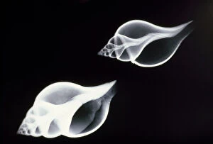
Background imageAnatomy Collection: Shells - x-ray

Shells - x-ray
PM-7161 SHELLS - X-RAY Pat Morris Please note that prints are for personal display purposes only and may not be reproduced in any way. contact details: prints@ardea.com tel: + 44 (0) 20 8318 1401

Background imageAnatomy Collection: Dog - Irish wolfhound, close-up of head

Dog - Irish wolfhound, close-up of head
LA-1571 Dog - Irish wolfhound, close-up of head Jean Michel Labat Please note that prints are for personal display purposes only and may not be reproduced in any way

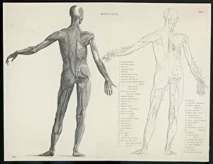
Background imageAnatomy Collection: Anatomy / Muscles

Anatomy / Muscles
First layer of muscles in the human body (back view)

Background imageAnatomy Collection: Anatomy / Sole of Foot

Anatomy / Sole of Foot
First and second stages of the dissection of the sole of the foot

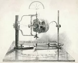
Background imageAnatomy Collection: Craniometer / Mid C19

Craniometer / Mid C19
A cranionmeter holds a skull in place

Background imageAnatomy Collection: Ideal Scientist

Ideal Scientist
The ideal scientist - interested in the old as well as the new, anatomy as well as astronomy, the laws of chance and of mathematics, the skill of chess, the hues of a rose

Background imageAnatomy Collection: Robert Burton

Robert Burton
ROBERT BURTON (1577-1640) English clergyman and author who wrote The Anatomy of Melancholy under the pseudonym of Democritus Junior in 1621

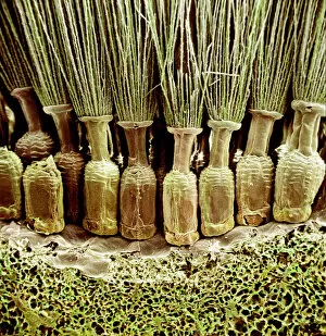
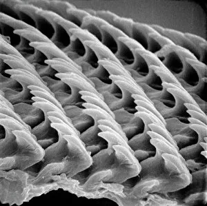
Background imageAnatomy Collection: Snail teeth

Snail teeth

Background imageAnatomy Collection: Medical / Anatomy / Eyes

Medical / Anatomy / Eyes
Medical/Anatomy/Eyes

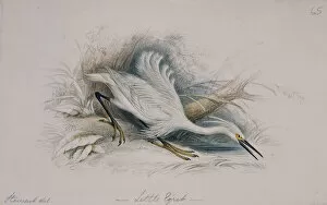
Background imageAnatomy Collection: Egretta garzetta, little egret

Egretta garzetta, little egret
A watercolour by James Hope Stewart, c. 1825-35. From Sir William Jardines The Naturalists Library

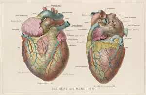
Background imageAnatomy Collection: German Heart

German Heart
Two views of the heart, with the parts labelled in German

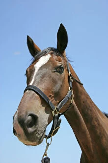
Background imageAnatomy Collection: Horse head. Nostril

Horse head. Nostril
JD-19370 Horse head. Nostril John Daniels Please note that prints are for personal display purposes only and may not be reproduced in any way

Background imageAnatomy Collection: Digestive Organs

Digestive Organs
Various diagrams depicting the digestive organs

Background imageAnatomy Collection: WHAT THE BRAIN DOES

WHAT THE BRAIN DOES
What the brain does

Background imageAnatomy Collection: Anatomy / Heart / Sibly

Anatomy / Heart / Sibly
Diagram of a human heart

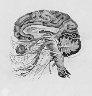
Background imageAnatomy Collection: Side View of Brain / C19

Side View of Brain / C19
A side view

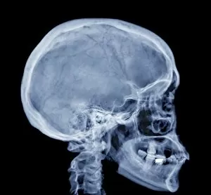
Background imageAnatomy Collection: Normal skull, X-ray

Normal skull, X-ray
Normal skull. Coloured X-ray of the head of a 28 year old patient

Background imageAnatomy Collection: Tarantula spider - face

Tarantula spider - face (Trochosa singoriensis). South Tuva, Russia


All Professionally Made to Order for Quick Shipping

"Unveiling the Intricacies of Anatomy: From Sensory Homunculus to Motor Homunculus" Step into the fascinating world of anatomy, where every detail tells a story. Explore the depths of human and animal structures as we delve into this captivating subject. Let's begin with Leonardo da Vinci's masterpiece, "Head of a Bear. " Created in 1480 but discovered in 1945, this artwork showcases da Vinci's unparalleled ability to capture anatomical accuracy. The intricate details reveal his keen observation skills and deep understanding of form. Moving on to our furry friends, the greyhound takes center stage. Discovering the anatomy of these majestic creatures unveils their remarkable speed and agility. From their sleek skeleton to their muscular build, it is evident why they are renowned for their racing abilities. But what about us humans? Enter the sensory homunculus – a visual representation that depicts how our brain perceives different body parts based on sensitivity levels. This map reveals intriguing insights into how our senses are distributed throughout our bodies. X-ray images provide another dimension to understanding anatomy. Take a glimpse at normal knees through an X-ray lens – marvel at the complexity hidden beneath our skin while appreciating its delicate balance between strength and flexibility. The backbone is often considered one of nature's most ingenious designs. Our human backbone includes ribs and pelvis, providing stability while allowing movement – truly an architectural marvel worth exploring further. Venturing deeper within ourselves, let us explore the intricacies of the human brain from an inferior view. Witness its complex network of connections responsible for controlling various bodily functions - truly awe-inspiring. As we journey back in time, we encounter hominid crania – remnants that shed light on our evolutionary history. These ancient skulls offer glimpses into early forms such as Australopithecus afarensis (AL 288-1), famously known as Lucy - bridging gaps between past and present.