Fraud Blocker Skip to main content

Anatomy Collection (page 8)

"Unveiling the Intricacies of Anatomy: From Sensory Homunculus to Motor Homunculus" Step into the fascinating world of anatomy, where every detail tells a story

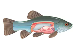
Background imageAnatomy Collection: Digital cross section illustration of fish showing gas bladder which contributes to the ability to

Digital cross section illustration of fish showing gas bladder which contributes to the ability to control buoyancy

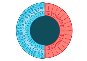
Background imageAnatomy Collection: Digital illustration of dilated pupil of human eye with contraction of outer radial muscle fibres

Digital illustration of dilated pupil of human eye with contraction of outer radial muscle fibres

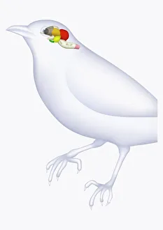
Background imageAnatomy Collection: Illustration of bird brain, including cerebrum, cerebellum, and medulla

Illustration of bird brain, including cerebrum, cerebellum, and medulla

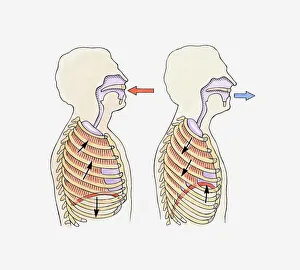
Background imageAnatomy Collection: Illustration showing diaphragm moving down when exhaling and up when inhaling

Illustration showing diaphragm moving down when exhaling and up when inhaling

Background imageAnatomy Collection: The science of human anatomy by Bartholomeo Eustachi

The science of human anatomy by Bartholomeo Eustachi, depicting the shape, size and relative position of the organs of the human body. Circa 18th century

Background imageAnatomy Collection: Human Musculature

Human Musculature
An anatomical diagram showing a rear view of the musculature of the upper human body, circa 1930. (Photo by Hulton Archive/Getty Images)

Background imageAnatomy Collection: Picture No. 10870743

Picture No. 10870743
Mountain Gorilla Skull - holotype 1902, Museum fur Natuurkunde, Berlin (Gorilla g. beringei) Date:

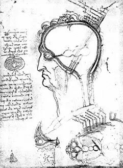
Background imageAnatomy Collection: Longitudinal section of an onion and sections of the human head

Longitudinal section of an onion and sections of the human head, eye and orbit: drawing by Leonardo da Vinci
LEONARDO: BRAIN, c1490. Longitudinal section of an onion and sections of the human head, eye and orbit: drawing by Leonardo da Vinci

Background imageAnatomy Collection: Model of branches of bronchial tree

Model of branches of bronchial tree

Background imageAnatomy Collection: Fulgora laternaria, peanut head bug

Fulgora laternaria, peanut head bug. How the peanut head bug got its name is self-evident. Its spectacular head is shaped like a peanut and, at six centimetres or so, is almost as long as its body

Background imageAnatomy Collection: Alcedo athhis, common kingfisher

Alcedo athhis, common kingfisher
Watercolour by Charles F. Tunnicliffe (c. 1973)

Background imageAnatomy Collection: Ara ararauna, blue-and-yellow maccaw

Ara ararauna, blue-and-yellow maccaw
Plate from William Hayes Portraits of Rare and Curious Birds, With Their Descriptions, From the Menagery of Osterley Park, London, (1794)

Background imageAnatomy Collection: Dendrocopos major, great spotted woodpecker

Dendrocopos major, great spotted woodpecker
Plate 26 from William MacGillivrays Watercolour drawings of British Animals (1831-1841)

Background imageAnatomy Collection: Passer domesticus, house sparrow

Passer domesticus, house sparrow
Plate 32 from John Goulds The Birds of Great Britain, Vol. 3 (1873). Hand coloured lithograph

Background imageAnatomy Collection: Podiceps cristatus, great crested grebe

Podiceps cristatus, great crested grebe
Plate 38 from John Goulds The Birds of Great Britain, Vol. 5 (1873). Hand coloured lithograph

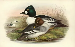
Background imageAnatomy Collection: Bucephala clangula, common goldeneye

Bucephala clangula, common goldeneye
Plate 31 from John Goulds The Birds of Great Britain, Vol. 5 (1873). Hand coloured lithograph

Background imageAnatomy Collection: Motacilla flava thunbergi, yellow wagtail

Motacilla flava thunbergi, yellow wagtail
Plate 4 from John Goulds The Birds of Great Britain, Vol. 3 (1873). Hand coloured lithograph

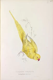
Background imageAnatomy Collection: Psittacula krameri manillensis, Indian ringneck parakeet (Lu

Psittacula krameri manillensis, Indian ringneck parakeet (Lu
Plate 33 from Edward Lears Illustrations of the Family of Psittacidae or Parrots (1832). Hand coloured lithograph

Background imageAnatomy Collection: Megalaima zeylanica, brown-headed barbet

Megalaima zeylanica, brown-headed barbet
Plate 28, painting by Pieter Cornelius de Bevere, from the Loten Collection of coloured drawings of Birds, Mammals, Insects & Plants, (1754-57)

Background imageAnatomy Collection: Loxodonta africana, African elephant

Loxodonta africana, African elephant
A close-up of an African elephant adult male bull, on display in the Mammal and Whale Gallery (number 24), at the Natural History Museum, London

Background imageAnatomy Collection: Semioptera wallacei, standardwing

Semioptera wallacei, standardwing
Plate 52, hand coloured lithograph by John and Elizabeth Gould from John Goulds The Birds of Australia, Supplementary volume, (1840-1848)

Background imageAnatomy Collection: Plecotus sp. long-eared bat

Plecotus sp. long-eared bat
A long-eared bat in flight (a microbat belonging to the Vespertilionidae family of vesper or evening bats). Photograph published in Bats by Phil Richardson, a Natural History Museum publication, 2002

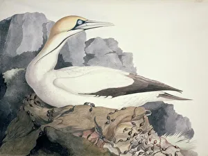
Background imageAnatomy Collection: Sula capensis, Cape Gannet

Sula capensis, Cape Gannet
Plate 2 from William MacGillivrays Watercolour drawings of British Animals (1831-1841)

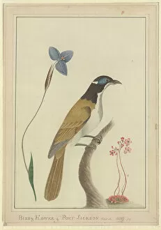
Background imageAnatomy Collection: Entomyzon cyanotis, blue-faced honeyeater

Entomyzon cyanotis, blue-faced honeyeater
Drawing No. 49, watercolour by George Raper (1792) from The Raper Collection

Background imageAnatomy Collection: Anopheles gambiae, mosquito

Anopheles gambiae, mosquito
Scanning electron microscope image showing a close-up of the compound eye of a female mosquito (x 2200 on a standard 9 cm wide print)

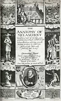
Background imageAnatomy Collection: Engraved title page

Engraved title page. From: The anatomy of melancholy. Date: 1632

Background imageAnatomy Collection: Modern Athenians plate 46 - Edinburgh

Modern Athenians plate 46 - Edinburgh
James Simson (d. 1876) doctor and president of the Royal College of Surgeons, approaching Alexander Monro (d. 1859) professor of Anatomy

Background imageAnatomy Collection: Well Rooted Woman

Well Rooted Woman
http://csaimages.com/images/istockprofile/csa_vector_dsp.jpg

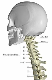
Background imageAnatomy Collection: anatomy, back bone, back view, bone, bone structure, bones, bones of the neck, cervical vertebrae

anatomy, back bone, back view, bone, bone structure, bones, bones of the neck, cervical vertebrae
anatomy, back bone, back view, bone, bone structure, bones, bones of t, Medical Image Collection, 87396753

Background imageAnatomy Collection: External Muscles of the Horse

External Muscles of the Horse

Background imageAnatomy Collection: Development of chicken egg anatomy engraving 1857

Development of chicken egg anatomy engraving 1857
Rank, johannes - The human being. 1 - 1894

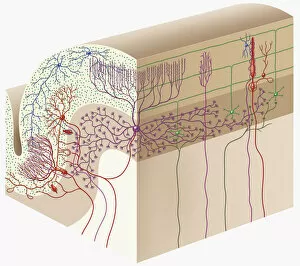
Background imageAnatomy Collection: Digital cross section illustration of human cerebellar cortex

Digital cross section illustration of human cerebellar cortex

Background imageAnatomy Collection: Digital illustration of direction of information in amygdala

Digital illustration of direction of information in amygdala

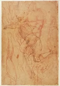
Background imageAnatomy Collection: Figure Study (red chalk on paper)

Figure Study (red chalk on paper)
BAL49879 Figure Study (red chalk on paper) by Buonarroti, Michelangelo (1475-1564); British Museum, London, UK; (add.info.: (W.6 verso); ); Italian, out of copyright

Background imageAnatomy Collection: Kodak jar with pebbles from Emperor Penguin (Aptenodytes for

Kodak jar with pebbles from Emperor Penguin (Aptenodytes for
Kodak jar with pebbles from emperor penguin (Aptenodytes forsteri) stomach Collected by Scotts British Antarctic Expedition 1910-1913, also known as the Terra Nova expedition

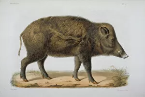
Background imageAnatomy Collection: Sus moupinensis, wild hog

Sus moupinensis, wild hog
Plate 80 from Recherches pour servir a l histoire naturelle des Mammifcres, Vols. 1-4, 1868-74, by Dr. Henri Milne-Edwards (1800-1885) & Dr. Alphonse Milne-Edwards (1835-1900)

Background imageAnatomy Collection: LS Plate 2 from the John Reeves Collection

LS Plate 2 from the John Reeves Collection
Large Series plate 2, a watercolour from the John Reeves Collection of Zoological Drawings from Canton, China

Background imageAnatomy Collection: Phalacrocorax carbo, great cormorant

Phalacrocorax carbo, great cormorant
Plate 52 from John Goulds The Birds of Great Britain, Vol. 5 (1873). Hand coloured lithograph

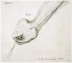
Background imageAnatomy Collection: Agkistrodon piscivorus, cottonmouth snake

Agkistrodon piscivorus, cottonmouth snake
Drawing 61 (Ewan 50) from the Botanical and zoological drawings (1756-1788) by William Bartram

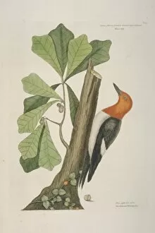
Background imageAnatomy Collection: Melanerpes erythrocephalus, red-headed woodpecker

Melanerpes erythrocephalus, red-headed woodpecker
Plate 20, hand coloured etching from The Natural History of Carolina, Florida and the Bahama islands (1731-43) Vol. 1 by Mark Catesby

Background imageAnatomy Collection: Plate 327 from The Gleanings of Natural History by George Ed

Plate 327 from The Gleanings of Natural History by George Ed
Plate 328, hand coloured copperplate etching from George Edwards The Gleanings of Natural History, Vol. 3 (1764). Annotated Brasilian saw-billed roller

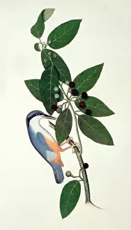
Background imageAnatomy Collection: Sitta sp. nuthatch

Sitta sp. nuthatch
Plate 40 from a bound volume called Indian Birds Colouredd. Artrist probably Sheikh Zayn al-Din, possibly compiled by Lady Impey

Background imageAnatomy Collection: Motacilla flava, yellow wagtail

Motacilla flava, yellow wagtail
Watercolour by Henrik Gr� ld (1924-5)

Background imageAnatomy Collection: Lynx sp. lynx

Lynx sp. lynx
Stuffed specimen from the collection of the Natural History Museum at Tring, part of the Natural History Museum, London

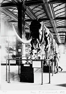
Background imageAnatomy Collection: Mastodon in Geological Gallery, December 1919

Mastodon in Geological Gallery, December 1919
The Guide Lecturer, John Henry Leonard, took this shot of two girls inspecting a primitive elephant or mastodon (Mammut americanus), in December 1919

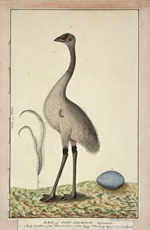
Background imageAnatomy Collection: Dromaius novaehollandiae, emu

Dromaius novaehollandiae, emu
Drawing No. 67, watercolour by George Raper (1792) from The Raper Collection

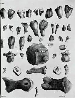
Background imageAnatomy Collection: Plate 17. Mantells Geology of Sussex

Plate 17. Mantells Geology of Sussex
Plate 17 from Illustrations of the Geology of Sussex, 1827 by G. A. Mantell


All Professionally Made to Order for Quick Shipping

"Unveiling the Intricacies of Anatomy: From Sensory Homunculus to Motor Homunculus" Step into the fascinating world of anatomy, where every detail tells a story. Explore the depths of human and animal structures as we delve into this captivating subject. Let's begin with Leonardo da Vinci's masterpiece, "Head of a Bear. " Created in 1480 but discovered in 1945, this artwork showcases da Vinci's unparalleled ability to capture anatomical accuracy. The intricate details reveal his keen observation skills and deep understanding of form. Moving on to our furry friends, the greyhound takes center stage. Discovering the anatomy of these majestic creatures unveils their remarkable speed and agility. From their sleek skeleton to their muscular build, it is evident why they are renowned for their racing abilities. But what about us humans? Enter the sensory homunculus – a visual representation that depicts how our brain perceives different body parts based on sensitivity levels. This map reveals intriguing insights into how our senses are distributed throughout our bodies. X-ray images provide another dimension to understanding anatomy. Take a glimpse at normal knees through an X-ray lens – marvel at the complexity hidden beneath our skin while appreciating its delicate balance between strength and flexibility. The backbone is often considered one of nature's most ingenious designs. Our human backbone includes ribs and pelvis, providing stability while allowing movement – truly an architectural marvel worth exploring further. Venturing deeper within ourselves, let us explore the intricacies of the human brain from an inferior view. Witness its complex network of connections responsible for controlling various bodily functions - truly awe-inspiring. As we journey back in time, we encounter hominid crania – remnants that shed light on our evolutionary history. These ancient skulls offer glimpses into early forms such as Australopithecus afarensis (AL 288-1), famously known as Lucy - bridging gaps between past and present.