Fraud Blocker Skip to main content

Anatomy Collection (page 12)

"Unveiling the Intricacies of Anatomy: From Sensory Homunculus to Motor Homunculus" Step into the fascinating world of anatomy, where every detail tells a story

Background imageAnatomy Collection: Anatomic plate in ' Anatomia Reformata' by Thomas Bartholin (1616-80) (Bartholinus)

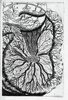
Anatomic plate in " Anatomia Reformata" by Thomas Bartholin (1616-80) (Bartholinus) from 1651 - Venas Lacteas
XEE5027928 Anatomic plate in " Anatomia Reformata" by Thomas Bartholin (1616-80) (Bartholinus) from 1651 - Venas Lacteas; (add.info)

Background imageAnatomy Collection: Frontispice de' De lactibus, sive, Lacteis venis, quarto vasorum mesaraicorum genere

Frontispice de" De lactibus, sive, Lacteis venis, quarto vasorum mesaraicorum genere, novo invento" de 1628
XEE5027828 Frontispice de" De lactibus, sive, Lacteis venis, quarto vasorum mesaraicorum genere, novo invento" de 1628

Background imageAnatomy Collection: Anatomic plate in 'Anatomia Reformata' by Thomas Bartholin (1616-80) (Bartholinus)

Anatomic plate in "Anatomia Reformata" by Thomas Bartholin (1616-80) (Bartholinus)
XEE5027942 Anatomic plate in "Anatomia Reformata" by Thomas Bartholin (1616-80) (Bartholinus) of 1651 - female reproductive organs; (add.info)

Background imageAnatomy Collection: Portrait of Albrecht von Haller (or Albert de Halle) (1708-1777) Swiss doctor, naturalist and poet

Portrait of Albrecht von Haller (or Albert de Halle) (1708-1777) Swiss doctor, naturalist and poet
ELD4907084 Portrait of Albrecht von Haller (or Albert de Halle) (1708-1777) Swiss doctor, naturalist and poet. 19th century (engraving); Private Collection; Photo © The Holbarn Archive.

Background imageAnatomy Collection: Portrait of Francois Achille Longet (1811-1871), French anatomist and physiologist

Portrait of Francois Achille Longet (1811-1871), French anatomist and physiologist. 19th century (engraving)
ELD4905408 Portrait of Francois Achille Longet (1811-1871), French anatomist and physiologist. 19th century (engraving); Private Collection; Photo © The Holbarn Archive.

Background imageAnatomy Collection: Title page of the anatomy ' Anatomia Humani Corporis' (1685) by Godefridi (Govert or Govard)

Title page of the anatomy " Anatomia Humani Corporis" (1685) by Godefridi (Govert or Govard)
PCT4281222 Title page of the anatomy " Anatomia Humani Corporis" (1685) by Godefridi (Govert or Govard) Bidloo (1649-1713) 18th century; (add.info)

Background imageAnatomy Collection: Internal muscles of the back, the deepest. Lithograph by Davesne, based on a drawing by Leveille

Internal muscles of the back, the deepest. Lithograph by Davesne, based on a drawing by Leveille
FLO4672659 Internal muscles of the back, the deepest. Lithograph by Davesne, based on a drawing by Leveille, in Petity Atlas complet d'Anatomie descriptive du Corps Humain, by Dr

Background imageAnatomy Collection: Frontal bone of the skull. Lithograph of Corbie, in Petity Atlas complet d'Anatomie descriptive du

Frontal bone of the skull. Lithograph of Corbie, in Petity Atlas complet d'Anatomie descriptive du Corps Humain
FLO4672402 Frontal bone of the skull. Lithograph of Corbie, in Petity Atlas complet d'Anatomie descriptive du Corps Humain, by Dr

Background imageAnatomy Collection: Muscles of the face, eyes and neck. Lithograph by Davesne, based on a drawing by Leveille

Muscles of the face, eyes and neck. Lithograph by Davesne, based on a drawing by Leveille
FLO4672617 Muscles of the face, eyes and neck. Lithograph by Davesne, based on a drawing by Leveille, in Petity Atlas complet d'Anatomie descriptive du Corps Humain, by Dr

Background imageAnatomy Collection: Olfactory and maxillary nerves. Lithograph by Davesne, based on a drawing by Leveille

Olfactory and maxillary nerves. Lithograph by Davesne, based on a drawing by Leveille
FLO4672503 Olfactory and maxillary nerves. Lithograph by Davesne, based on a drawing by Leveille, in Petity Atlas complet d'Anatomie descriptive du Corps Humain, by Dr

Background imageAnatomy Collection: Saphene vein and medial plantar nerves. Lithograph of Corbie, based on a drawing by Leveille

Saphene vein and medial plantar nerves. Lithograph of Corbie, based on a drawing by Leveille
FLO4672470 Saphene vein and medial plantar nerves. Lithograph of Corbie, based on a drawing by Leveille, in Petity Atlas complet d'Anatomie descriptive du Corps Humain, by Dr

Background imageAnatomy Collection: Skeleton of a crocodile, Crocodylus niloticus. Copperplate engraving by Heath after an

Skeleton of a crocodile, Crocodylus niloticus. Copperplate engraving by Heath after an illustration by George Shaw
FLO4694870 Skeleton of a crocodile, Crocodylus niloticus. Copperplate engraving by Heath after an illustration by George Shaw from his General Zoology, Amphibia, London

Background imageAnatomy Collection: Anatomy of human intestines from the front. Lithograph from Lorenz Oken's Universal Natural History

Anatomy of human intestines from the front. Lithograph from Lorenz Oken's Universal Natural History
FLO4684808 Anatomy of human intestines from the front. Lithograph from Lorenz Oken's Universal Natural History, Allgemeine Naturgeschichte fur alle Stande, Stuttgart

Background imageAnatomy Collection: Anatomy of the nervous system in the heart, neck and arm. Lithograph by C

Anatomy of the nervous system in the heart, neck and arm. Lithograph by C
FLO4684805 Anatomy of the nervous system in the heart, neck and arm. Lithograph by C. Schach from Lorenz Oken's Universal Natural History, Allgemeine Naturgeschichte fur alle Stande, Stuttgart

Background imageAnatomy Collection: Anatomy of the human skeleton, 1839 (lithograph)

Anatomy of the human skeleton, 1839 (lithograph)
FLO4684794 Anatomy of the human skeleton, 1839 (lithograph); (add.info.: Anatomy of the human skeleton. Lithograph from Lorenz Oken's Universal Natural History)

Background imageAnatomy Collection: Anatomy of the human ear and eye, 1839 (lithograph)

Anatomy of the human ear and eye, 1839 (lithograph)
FLO4684845 Anatomy of the human ear and eye, 1839 (lithograph); (add.info.: Anatomy of the human ear and eye. Lithograph from Lorenz Oken's Universal Natural History)

Background imageAnatomy Collection: Anatomy of the human venal system in the arms. Lithograph by C

Anatomy of the human venal system in the arms. Lithograph by C. Mayer from Lorenz Oken's Universal Natural History
FLO4684837 Anatomy of the human venal system in the arms. Lithograph by C. Mayer from Lorenz Oken's Universal Natural History, Allgemeine Naturgeschichte fur alle Stande, Stuttgart

Background imageAnatomy Collection: Jan Evangelista Purkyne, 19th century (engraving)

Jan Evangelista Purkyne, 19th century (engraving)
3733542 Jan Evangelista Purkyne, 19th century (engraving); (add.info.: Portrait of Jan Evangelista Purkyne (1787-1869) a Czech anatomist and physiologist

Background imageAnatomy Collection: Georges CUVIER (1769-1832), French anatomist and paleontologist

Georges CUVIER (1769-1832), French anatomist and paleontologist
JAB4121367 Georges CUVIER (1769-1832), French anatomist and paleontologist.; (add.info.: Georges CUVIER (1769-1832), French anatomist and paleontologist.); Photo ©Jaime Abecasis.

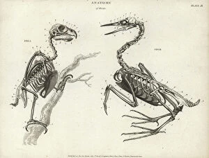
Background imageAnatomy Collection: Anatomy of birds: skeleton. Copperplate engraving by Milton from Abraham Rees' Cyclopedia or

Anatomy of birds: skeleton. Copperplate engraving by Milton from Abraham Rees' Cyclopedia or Universal Dictionary
FLO4593655 Anatomy of birds: skeleton. Copperplate engraving by Milton from Abraham Rees' Cyclopedia or Universal Dictionary of Arts, Sciences and Literature, Longman, Hurst, Rees

Background imageAnatomy Collection: Anatomy of human musculature from the back showing inner muscles, 1820 (copperplate engraving)

Anatomy of human musculature from the back showing inner muscles, 1820 (copperplate engraving)
FLO4591048 Anatomy of human musculature from the back showing inner muscles, 1820 (copperplate engraving); (add.info.: Anatomy of human musculature from the back showing inner muscles)

Background imageAnatomy Collection: Anatomy of human internal organs from the back showing spine, lungs, liver, intestines

Anatomy of human internal organs from the back showing spine, lungs, liver, intestines, gallbladder, etc
FLO4594255 Anatomy of human internal organs from the back showing spine, lungs, liver, intestines, gallbladder, etc. Copperplate engraving by Milton from Abraham Rees' Cyclopedia or Universal

Background imageAnatomy Collection: Anatomy of the horse: musculature. Copperplate engraving by Milton from Abraham Rees' Cyclopedia

Anatomy of the horse: musculature. Copperplate engraving by Milton from Abraham Rees' Cyclopedia or Universal
FLO4593561 Anatomy of the horse: musculature. Copperplate engraving by Milton from Abraham Rees' Cyclopedia or Universal Dictionary of Arts, Sciences and Literature, Longman, Hurst, Rees

Background imageAnatomy Collection: Anatomy of the human ear showing cochlea and internal parts. Copperplate engraving by Milton

Anatomy of the human ear showing cochlea and internal parts. Copperplate engraving by Milton from Abraham Rees'
FLO4591625 Anatomy of the human ear showing cochlea and internal parts. Copperplate engraving by Milton from Abraham Rees' Cyclopedia or Universal Dictionary of Arts, Sciences and Literature

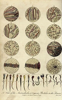
Background imageAnatomy Collection: Microscopic views of human spermatozoa. View of the animalculae or organic particles in the semen

Microscopic views of human spermatozoa. View of the animalculae or organic particles in the semen
FLO4714918 Microscopic views of human spermatozoa. View of the animalculae or organic particles in the semen. Handcoloured copperplate engraving by J

Background imageAnatomy Collection: Teething, Growth - Normal Life & Health by J. Rengade.Drawing A. Demarle 1881

Teething, Growth - Normal Life & Health by J. Rengade.Drawing A. Demarle 1881
JAB4120225 Teething, Growth - Normal Life & Health by J. Rengade.Drawing A. Demarle 1881; (add.info.: Teething, Growth - Normal Life & Health by J. Rengade.Drawing A)

Background imageAnatomy Collection: Hip muscles - 1866 illustration showing the muscles (brown), tendons (white) and bones (beige)

Hip muscles - 1866 illustration showing the muscles (brown), tendons (white) and bones (beige) of the human hip (centre)
JAB4119507 Hip muscles - 1866 illustration showing the muscles (brown), tendons (white) and bones (beige) of the human hip (centre) - Seen here are: Fig 1: sacrum and coccyx (tail bone); Fig 2

Background imageAnatomy Collection: Chest and neck muscles - 1866 anatomical illustration showing the structure of the muscles (brown)

Chest and neck muscles - 1866 anatomical illustration showing the structure of the muscles (brown) and tendons (white)
JAB4119483 Chest and neck muscles - 1866 anatomical illustration showing the structure of the muscles (brown) and tendons (white) found in the rear of the human head (top) and neck (upper centre)

Background imageAnatomy Collection: Pelvic and abdominal muscles - 1866 illustration of the internal muscles (brown) and tendons (white)

Pelvic and abdominal muscles - 1866 illustration of the internal muscles (brown) and tendons (white)
JAB4119447 Pelvic and abdominal muscles - 1866 illustration of the internal muscles (brown) and tendons (white) around the human pelvis (lower centre) and abdomen

Background imageAnatomy Collection: Rib cage anatomy - 1866 illustration of the muscles (brown), tendons (white) and bones (beige)

Rib cage anatomy - 1866 illustration of the muscles (brown), tendons (white) and bones (beige) of the human rib cage
JAB4119446 Rib cage anatomy - 1866 illustration of the muscles (brown), tendons (white) and bones (beige) of the human rib cage, showing the sternum (breast bone, Fig 1), ribs

Background imageAnatomy Collection: Leg and knee muscles - 1866 anatomical illustration showing the muscles (brown) and tendons (white)

Leg and knee muscles - 1866 anatomical illustration showing the muscles (brown) and tendons (white)
JAB4119438 Leg and knee muscles - 1866 anatomical illustration showing the muscles (brown) and tendons (white) of the human leg around the knee (centre) - Outer area of the knee - Vast inner muscle

Background imageAnatomy Collection: Diaphragm anatomy - 1866 anatomical illustration showing the human diaphragm (top: centre)

Diaphragm anatomy - 1866 anatomical illustration showing the human diaphragm (top: centre)
JAB4119430 Diaphragm anatomy - 1866 anatomical illustration showing the human diaphragm (top: centre, bottom: upper-centre)

Background imageAnatomy Collection: Arm muscles - 1866 illustration showing the structure of the muscles (brown) and tendons (white)

Arm muscles - 1866 illustration showing the structure of the muscles (brown) and tendons (white)
JAB4119402 Arm muscles - 1866 illustration showing the structure of the muscles (brown) and tendons (white) of the human arm - The internal

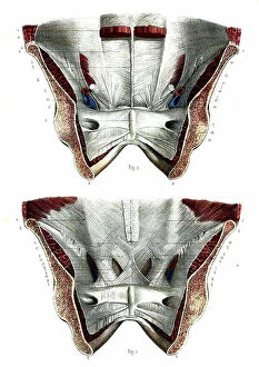
Background imageAnatomy Collection: Pelvis anatomy - 1866 illustration showing the anatomy of the male (top) and female (bottom)

Pelvis anatomy - 1866 illustration showing the anatomy of the male (top) and female (bottom)
JAB4119375 Pelvis anatomy - 1866 illustration showing the anatomy of the male (top) and female (bottom) pelvis - Aponevroses abdominales, posterior plane - Fig 1: Crete iliaque Fig 2: Crete iliaque

Background imageAnatomy Collection: Human hand - 1866 anatomical illustration showing the muscles (brown) and tendons (white)

Human hand - 1866 anatomical illustration showing the muscles (brown) and tendons (white) of the upper (right)
JAB4119374 Human hand - 1866 anatomical illustration showing the muscles (brown) and tendons (white) of the upper (right) and lower (left)

Background imageAnatomy Collection: Lung anatomy 1866 illustrations showing the external anatomy of the human lungs (beige)

Lung anatomy 1866 illustrations showing the external anatomy of the human lungs (beige)
JAB4119246 Lung anatomy 1866 illustrations showing the external anatomy of the human lungs (beige), with the bronchi and trachea (white)

Background imageAnatomy Collection: Female urogenital system 1866 illustration of a section through the urogenital system of a human

Female urogenital system 1866 illustration of a section through the urogenital system of a human female Anatomy
JAB4119243 Female urogenital system 1866 illustration of a section through the urogenital system of a human female Anatomy, l'uretre - Descriptive Anatomy

Background imageAnatomy Collection: Male perineum 1866 illustration showing the muscles and other structures of the male perineum

Male perineum 1866 illustration showing the muscles and other structures of the male perineum - the region of the body
JAB4119197 Male perineum 1866 illustration showing the muscles and other structures of the male perineum - the region of the body located between the scrotum and anus Le perinee de l'homme

Background imageAnatomy Collection: Prostate gland 1866 illustrations showing the structure of the prostate gland Prostate structure

Prostate gland 1866 illustrations showing the structure of the prostate gland Prostate structure - Fig 1
JAB4119151 Prostate gland 1866 illustrations showing the structure of the prostate gland Prostate structure - Fig 1: Prostate length cut Fig 2: Middle section cut

Background imageAnatomy Collection: Skeleton of an extinct pterodactyl. Lithograph by St. Schillinger from Lorenz Oken's Universal

Skeleton of an extinct pterodactyl. Lithograph by St. Schillinger from Lorenz Oken's Universal Natural History
FLO4684698 Skeleton of an extinct pterodactyl. Lithograph by St. Schillinger from Lorenz Oken's Universal Natural History, Allgemeine Naturgeschichte fur alle Stande, Stuttgart

Background imageAnatomy Collection: Brain sections, cerebellum and ventricles. Lithograph by Davesne, based on a drawing by Leveille

Brain sections, cerebellum and ventricles. Lithograph by Davesne, based on a drawing by Leveille
FLO4673145 Brain sections, cerebellum and ventricles. Lithograph by Davesne, based on a drawing by Leveille, in Petity Atlas complet d'Anatomie descriptive du Corps Humain, by Dr

Background imageAnatomy Collection: Cross sections of the brain. Lithograph of Corbie, based on a drawing by Leveille

Cross sections of the brain. Lithograph of Corbie, based on a drawing by Leveille
FLO4673108 Cross sections of the brain. Lithograph of Corbie, based on a drawing by Leveille, in Petity Atlas complet d'Anatomie descriptive du Corps Humain, by Dr

Background imageAnatomy Collection: Lymphatic system of the abdomen. Lithograph of Corbie, based on a drawing by Leveille

Lymphatic system of the abdomen. Lithograph of Corbie, based on a drawing by Leveille
FLO4673043 Lymphatic system of the abdomen. Lithograph of Corbie, based on a drawing by Leveille, in Petity Atlas complet d'Anatomie descriptive du Corps Humain, by Dr

Background imageAnatomy Collection: Pelvis and arm circulatory system. Lithograph by Davesne, based on a drawing by Leveille

Pelvis and arm circulatory system. Lithograph by Davesne, based on a drawing by Leveille
FLO4672946 Pelvis and arm circulatory system. Lithograph by Davesne, based on a drawing by Leveille, in Petity Atlas complet d'Anatomie descriptive du Corps Humain, by Dr

Background imageAnatomy Collection: Circulatory system of the abdomen. Lithograph of Corbie, based on a drawing by Leveille

Circulatory system of the abdomen. Lithograph of Corbie, based on a drawing by Leveille
FLO4672925 Circulatory system of the abdomen. Lithograph of Corbie, based on a drawing by Leveille, in Petity Atlas complet d'Anatomie descriptive du Corps Humain, by Dr

Background imageAnatomy Collection: The circulatory system of the brain. Lithograph of Choubard, based on a drawing by Leveille

The circulatory system of the brain. Lithograph of Choubard, based on a drawing by Leveille
FLO4672884 The circulatory system of the brain. Lithograph of Choubard, based on a drawing by Leveille, in Petity Atlas complet d'Anatomie descriptive du Corps Humain, by Dr

Background imageAnatomy Collection: Sections of the heart. Lithograph of Corbie, based on a drawing by Leveille

Sections of the heart. Lithograph of Corbie, based on a drawing by Leveille
FLO4672832 Sections of the heart. Lithograph of Corbie, based on a drawing by Leveille, in Petity Atlas complet d'Anatomie descriptive du Corps Humain, by Dr

Background imageAnatomy Collection: Human female skeleton from the front, with ostrich skeleton by William Cheselden

Human female skeleton from the front, with ostrich skeleton by William Cheselden
FLO4565814 Human female skeleton from the front, with ostrich skeleton by William Cheselden. Copperplate engraving by Edward Mitchell after an anatomical illustration by Jean-Joseph Sue from John


All Professionally Made to Order for Quick Shipping

"Unveiling the Intricacies of Anatomy: From Sensory Homunculus to Motor Homunculus" Step into the fascinating world of anatomy, where every detail tells a story. Explore the depths of human and animal structures as we delve into this captivating subject. Let's begin with Leonardo da Vinci's masterpiece, "Head of a Bear. " Created in 1480 but discovered in 1945, this artwork showcases da Vinci's unparalleled ability to capture anatomical accuracy. The intricate details reveal his keen observation skills and deep understanding of form. Moving on to our furry friends, the greyhound takes center stage. Discovering the anatomy of these majestic creatures unveils their remarkable speed and agility. From their sleek skeleton to their muscular build, it is evident why they are renowned for their racing abilities. But what about us humans? Enter the sensory homunculus – a visual representation that depicts how our brain perceives different body parts based on sensitivity levels. This map reveals intriguing insights into how our senses are distributed throughout our bodies. X-ray images provide another dimension to understanding anatomy. Take a glimpse at normal knees through an X-ray lens – marvel at the complexity hidden beneath our skin while appreciating its delicate balance between strength and flexibility. The backbone is often considered one of nature's most ingenious designs. Our human backbone includes ribs and pelvis, providing stability while allowing movement – truly an architectural marvel worth exploring further. Venturing deeper within ourselves, let us explore the intricacies of the human brain from an inferior view. Witness its complex network of connections responsible for controlling various bodily functions - truly awe-inspiring. As we journey back in time, we encounter hominid crania – remnants that shed light on our evolutionary history. These ancient skulls offer glimpses into early forms such as Australopithecus afarensis (AL 288-1), famously known as Lucy - bridging gaps between past and present.