Fraud Blocker Skip to main content

Anatomy Collection (page 14)

"Unveiling the Intricacies of Anatomy: From Sensory Homunculus to Motor Homunculus" Step into the fascinating world of anatomy, where every detail tells a story

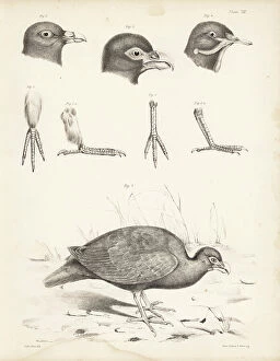
Background imageAnatomy Collection: Cranium 1-4 and part of sternum 5,6 of the extinct Rodrigues solitaire

Cranium 1-4 and part of sternum 5,6 of the extinct Rodrigues solitaire. Lithograph by Joseph Dinkel after Werner
FLO4612182 Cranium 1-4 and part of sternum 5,6 of the extinct Rodrigues solitaire. Lithograph by Joseph Dinkel after Werner from Hugh Edwin Strickland and Alexander Gordon Melville's The Dodo

Background imageAnatomy Collection: Skulls of tooth-billed pigeon, Didunculus strigirostris, critically endangered 1,7,8,9,10 dodo

Skulls of tooth-billed pigeon, Didunculus strigirostris, critically endangered 1,7,8,9,10 dodo, Raphus cucullatus 2
FLO4612171 Skulls of tooth-billed pigeon, Didunculus strigirostris, critically endangered 1,7,8,9,10 dodo, Raphus cucullatus 2, yellow-footed green pigeon, Treron chlorigaster 3,6, crowned pigeon

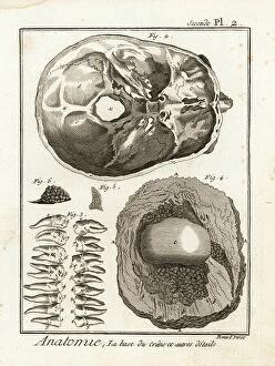
Background imageAnatomy Collection: Base of the skull, human teeth, knee cap (after Clopton Havers), mucus gland, etc

Base of the skull, human teeth, knee cap (after Clopton Havers), mucus gland, etc
FLO4611538 Base of the skull, human teeth, knee cap (after Clopton Havers), mucus gland, etc. Copperplate engraving by Robert Benard from Denis Diderot's Encyclopedia, Pellet, Geneva

Background imageAnatomy Collection: Interior of the brain and cerebellum. Copperplate engraving by Robert Benard after an illustration

Interior of the brain and cerebellum. Copperplate engraving by Robert Benard after an illustration by Albrecht von
FLO4611652 Interior of the brain and cerebellum. Copperplate engraving by Robert Benard after an illustration by Albrecht von Haller from Denis Diderot's Encyclopedia, Pellet, Geneva

Background imageAnatomy Collection: Arteries in the face. Copperplate engraving by Robert Benard after an illustration by Albrecht von

Arteries in the face. Copperplate engraving by Robert Benard after an illustration by Albrecht von Haller from Denis
FLO4611618 Arteries in the face. Copperplate engraving by Robert Benard after an illustration by Albrecht von Haller from Denis Diderot's Encyclopedia, Pellet, Geneva

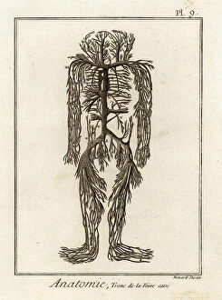
Background imageAnatomy Collection: Venal system from the vena cava in the human body. Copperplate engraving by Robert Benard after an

Venal system from the vena cava in the human body. Copperplate engraving by Robert Benard after an illustration by
FLO4611599 Venal system from the vena cava in the human body. Copperplate engraving by Robert Benard after an illustration by James Drake from Denis Diderot's Encyclopedia, Pellet, Geneva

Background imageAnatomy Collection: Details of arteries, veins, lymph acorns, etc. Copperplate engraving by Robert Benard after

Details of arteries, veins, lymph acorns, etc. Copperplate engraving by Robert Benard after from Denis Diderot's
FLO4611594 Details of arteries, veins, lymph acorns, etc. Copperplate engraving by Robert Benard after from Denis Diderot's Encyclopedia, Pellet, Geneva, 1779.; (add.info.: Details of arteries)

Background imageAnatomy Collection: Upper and lower views of the skull of a dodo. Lithograph from Hugh Edwin Strickland

Upper and lower views of the skull of a dodo. Lithograph from Hugh Edwin Strickland
FLO4612153 Upper and lower views of the skull of a dodo. Lithograph from Hugh Edwin Strickland and Alexander Gordon Melville's The Dodo and its Kindred, London, Reeve, Benham and Reeve

Background imageAnatomy Collection: Tooth billed pigeon, Didunculus strigirostris 1,2 (critically endangered), Bruce's green pigeon

Tooth billed pigeon, Didunculus strigirostris 1,2 (critically endangered), Bruce's green pigeon
FLO4612139 Tooth billed pigeon, Didunculus strigirostris 1,2 (critically endangered), Bruce's green pigeon, Treron waalia (Treron abyssinica 3,5) and Levaillant's dove

Background imageAnatomy Collection: Jean Louis Petite's machine to compress femoral artere during thigh amputation, bandage

Jean Louis Petite's machine to compress femoral artere during thigh amputation, bandage
FLO4611844 Jean Louis Petite's machine to compress femoral artere during thigh amputation, bandage, aneurysm needle and scissors - Plate taken from " L'Encyclopedie" by Denis

Background imageAnatomy Collection: Anatomy of the horse: stomach and intestines. Copperplate engraving by Scott from Abraham Rees'

Anatomy of the horse: stomach and intestines. Copperplate engraving by Scott from Abraham Rees' Cyclopedia or
FLO4593968 Anatomy of the horse: stomach and intestines. Copperplate engraving by Scott from Abraham Rees' Cyclopedia or Universal Dictionary of Arts, Sciences and Literature, Longman, Hurst

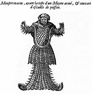
Background imageAnatomy Collection: Engraving of the book of Ambroise Pare: ' Monsters and Prodiges' (first edition 1573)

Engraving of the book of Ambroise Pare: " Monsters and Prodiges" (first edition 1573)
PCT4260964 Engraving of the book of Ambroise Pare: " Monsters and Prodiges" (first edition 1573, appendix: marine monsters)

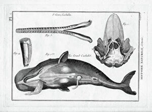
Background imageAnatomy Collection: Le grand sperm whale in ' Painting Encyclopedique et Methodique des trois Regnes de la Nature' by

Le grand sperm whale in " Painting Encyclopedique et Methodique des trois Regnes de la Nature" by Pierre
FAT4299958 Le grand sperm whale in " Painting Encyclopedique et Methodique des trois Regnes de la Nature" by Pierre Joseph Bonnaterre (1762-1804), Paris

Background imageAnatomy Collection: Skeleton of a European perch, Perca fluviatilis, from Dr. Barclay's Museum, Edinburgh

Skeleton of a European perch, Perca fluviatilis, from Dr. Barclay's Museum, Edinburgh
FLO4565818 Skeleton of a European perch, Perca fluviatilis, from Dr. Barclay's Museum, Edinburgh. Copperplate engraving by Edward Mitchell after an anatomical illustration by Robert Kaye Greville

Background imageAnatomy Collection: Rear view of the human trunk skeleton including spine, ribs and pelvis

Rear view of the human trunk skeleton including spine, ribs and pelvis. Copperplate engraving by Edward Mitchell after
FLO4565792 Rear view of the human trunk skeleton including spine, ribs and pelvis. Copperplate engraving by Edward Mitchell after an anatomical illustration by Jean-Joseph Sue from John Barclay's

Background imageAnatomy Collection: Views of the bones in the human foot from ankle to toe. Copperplate engraving by Edward Mitchell

Views of the bones in the human foot from ankle to toe. Copperplate engraving by Edward Mitchell after an anatomical
FLO4565738 Views of the bones in the human foot from ankle to toe. Copperplate engraving by Edward Mitchell after an anatomical illustration by Jean-Joseph Sue from John Barclay's A Series of

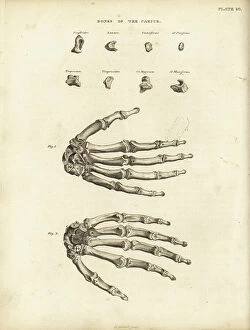
Background imageAnatomy Collection: Bones of the carpus, metacarpus and fingers in the human hand

Bones of the carpus, metacarpus and fingers in the human hand. Copperplate engraving by Edward Mitchell after an
FLO4565722 Bones of the carpus, metacarpus and fingers in the human hand. Copperplate engraving by Edward Mitchell after an anatomical illustration from John Barclay's A Series of Engravings of

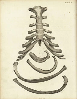
Background imageAnatomy Collection: Sternum and cartilage of the human ribcage, with several ribs

Sternum and cartilage of the human ribcage, with several ribs. Copperplate engraving by Edward Mitchell after an
FLO4565702 Sternum and cartilage of the human ribcage, with several ribs. Copperplate engraving by Edward Mitchell after an anatomical illustration by Jean-Joseph Sue from John Barclay's A Series

Background imageAnatomy Collection: Ribs and sternum in the human ribcage. Copperplate engraving by Edward Mitchell after an

Ribs and sternum in the human ribcage. Copperplate engraving by Edward Mitchell after an anatomical illustration by
FLO4565699 Ribs and sternum in the human ribcage. Copperplate engraving by Edward Mitchell after an anatomical illustration by Jean-Joseph Sue from John Barclay's A Series of Engravings of

Background imageAnatomy Collection: Front view of the human trunk skeleton including spine, ribs and pelvis

Front view of the human trunk skeleton including spine, ribs and pelvis. Copperplate engraving by Edward Mitchell after
FLO4565690 Front view of the human trunk skeleton including spine, ribs and pelvis. Copperplate engraving by Edward Mitchell after an anatomical illustration by Jean-Joseph Suefrom John Barclay's

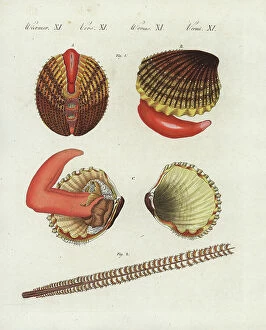
Background imageAnatomy Collection: Prickly cockle - bucarde - Acanthocardia echinata (Cardium echinatum) 1

Prickly cockle - bucarde - Acanthocardia echinata (Cardium echinatum) 1, and slender sea pen - plume elegante
FLO4576265 Prickly cockle - bucarde - Acanthocardia echinata (Cardium echinatum) 1, and slender sea pen - plume elegante - Virgularia mirabilis (Pennatula mirabilis) 2

Background imageAnatomy Collection: Adult crane desarticule 1867

Adult crane desarticule 1867
JAB4120023 Adult crane desarticule 1867; (add.info.: Adult crane desarticule 1867); Photo ©Jaime Abecasis.

Background imageAnatomy Collection: Bladder anatomy 1866 illustrations showing the internal (top) and external (bottom)

Bladder anatomy 1866 illustrations showing the internal (top) and external (bottom)
JAB4119242 Bladder anatomy 1866 illustrations showing the internal (top) and external (bottom) anatomy of the male urinary bladder, showing its muscular coating (brown)

Background imageAnatomy Collection: Voice box and thyroid 1866 illustration showing the anatomy of the larynx (voice box)

Voice box and thyroid 1866 illustration showing the anatomy of the larynx (voice box) and thyroid (brown)
JAB4119213 Voice box and thyroid 1866 illustration showing the anatomy of the larynx (voice box) and thyroid (brown, lower centre) Shown here are the hyoid bone (A), thyroid cartilage (B)

Background imageAnatomy Collection: Chest muscles - 1866 illustration showing the structure of the muscles (brown) and tendons (white)

Chest muscles - 1866 illustration showing the structure of the muscles (brown) and tendons (white) of the human chest
JAB4119479 Chest muscles - 1866 illustration showing the structure of the muscles (brown) and tendons (white) of the human chest

Background imageAnatomy Collection: Leg and foot muscles - 1866 anatomical illustration showing the muscles (brown) and tendons (white)

Leg and foot muscles - 1866 anatomical illustration showing the muscles (brown) and tendons (white)
JAB4119476 Leg and foot muscles - 1866 anatomical illustration showing the muscles (brown) and tendons (white) of the right foot (left)

Background imageAnatomy Collection: Shoulder muscles - 1866 illustration of the muscles (brown) and tendons (white)

Shoulder muscles - 1866 illustration of the muscles (brown) and tendons (white)
JAB4119462 Shoulder muscles - 1866 illustration of the muscles (brown) and tendons (white) associated with the human collarbone (clavicle, top) and shoulder blade (scapula)

Background imageAnatomy Collection: Chest and shoulder muscles - 1866 anatomical illustration showing the structure of the muscles

Chest and shoulder muscles - 1866 anatomical illustration showing the structure of the muscles (brown)
JAB4119433 Chest and shoulder muscles - 1866 anatomical illustration showing the structure of the muscles (brown) and tendons (white)

Background imageAnatomy Collection: Hand muscles - 1866 anatomical illustration showing the muscles (brown) and tendons (white)

Hand muscles - 1866 anatomical illustration showing the muscles (brown) and tendons (white) of the human hand
JAB4119426 Hand muscles - 1866 anatomical illustration showing the muscles (brown) and tendons (white) of the human hand, with insets (left)

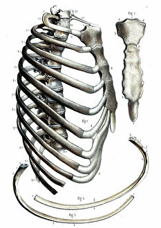
Background imageAnatomy Collection: Rib cage anatomy - 1866 illustration showing the bones of the human rib cage

Rib cage anatomy - 1866 illustration showing the bones of the human rib cage, including the ribs (bottom)
JAB4119411 Rib cage anatomy - 1866 illustration showing the bones of the human rib cage, including the ribs (bottom), spine (centre-left) and sternum (breast bone, centre and upper right) Le Thorax

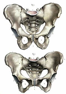
Background imageAnatomy Collection: Pelvis anatomy - 1866 illustration showing the anatomy of the male (top) and female (bottom)

Pelvis anatomy - 1866 illustration showing the anatomy of the male (top) and female (bottom) pelvis - The pelvis
JAB4119404 Pelvis anatomy - 1866 illustration showing the anatomy of the male (top) and female (bottom) pelvis - The pelvis, anatomy, osteology - Fig 1: female pelvis Fig 2: male pelvis, Musculotion

Background imageAnatomy Collection: Hip muscles - 1866 illustration showing the structure of the muscles (brown) and tendons (white)

Hip muscles - 1866 illustration showing the structure of the muscles (brown) and tendons (white)
JAB4119401 Hip muscles - 1866 illustration showing the structure of the muscles (brown) and tendons (white) of the upper right leg and hip

Background imageAnatomy Collection: Arm muscles - 1866 illustration showing the structure of the muscles (brown) and tendons (white)

Arm muscles - 1866 illustration showing the structure of the muscles (brown) and tendons (white)
JAB4119391 Arm muscles - 1866 illustration showing the structure of the muscles (brown) and tendons (white) of the human arm and top of the hand (bottom left and right)

Background imageAnatomy Collection: Abdominal muscles - 1866 illustration showing the structure of the muscles (brown)

Abdominal muscles - 1866 illustration showing the structure of the muscles (brown) and tendons (white)
JAB4119387 Abdominal muscles - 1866 illustration showing the structure of the muscles (brown) and tendons (white) found in the rear of the human trunk - L'abdomen - Anatomy - Region anterieure de

Background imageAnatomy Collection: Leg muscles - 1866 illustration showing the structure of the muscles (brown) and tendons (white)

Leg muscles - 1866 illustration showing the structure of the muscles (brown) and tendons (white)
JAB4119363 Leg muscles - 1866 illustration showing the structure of the muscles (brown) and tendons (white) of the rear of the lower leg - Anatomy du dos de la leg - Fig 1 et 2

Background imageAnatomy Collection: Andreas Vesalius

Andreas Vesalius
3586025 Andreas Vesalius; (add.info.: Portrait of Andreas Vesalius (1514-1564) a Flemish/Netherlandish anatomist, physician, and author of books on human anatomy

Background imageAnatomy Collection: Anatomy of the human eye, showing eyeball, optic nerve, muscle, cornea, retina, sections through eye

Anatomy of the human eye, showing eyeball, optic nerve, muscle, cornea, retina, sections through eye
FLO4591961 Anatomy of the human eye, showing eyeball, optic nerve, muscle, cornea, retina, sections through eye. Copperplate engraving by Milton from Abraham Rees' Cyclopedia or Universal

Background imageAnatomy Collection: Anatomy of human internal organs from the back showing stomach, liver, intestines, gallbladder, etc

Anatomy of human internal organs from the back showing stomach, liver, intestines, gallbladder, etc
FLO4590759 Anatomy of human internal organs from the back showing stomach, liver, intestines, gallbladder, etc. Copperplate engraving by Milton from Abraham Rees' Cyclopedia or Universal

Background imageAnatomy Collection: Anatomy of human musculature showing outer muscles from the front, 1820 (copperplate engraving)

Anatomy of human musculature showing outer muscles from the front, 1820 (copperplate engraving)
FLO4591432 Anatomy of human musculature showing outer muscles from the front, 1820 (copperplate engraving); (add.info.: Anatomy of human musculature showing outer muscles from the front)

Background imageAnatomy Collection: Anatomy of human musculature from the back showing outer muscles, 1820 (copperplate engraving)

Anatomy of human musculature from the back showing outer muscles, 1820 (copperplate engraving)
FLO4591171 Anatomy of human musculature from the back showing outer muscles, 1820 (copperplate engraving); (add.info.: Anatomy of human musculature from the back showing outer muscles)

Background imageAnatomy Collection: Anatomy of human internal organs from the front showing heart, stomach, intestines

Anatomy of human internal organs from the front showing heart, stomach, intestines, kidneys and bladder
FLO4590679 Anatomy of human internal organs from the front showing heart, stomach, intestines, kidneys and bladder. Copperplate engraving by Milton from Abraham Rees' Cyclopedia or Universal

Background imageAnatomy Collection: Head muscles. Lithograph of Lebrun, based on a drawing by Leveille

Head muscles. Lithograph of Lebrun, based on a drawing by Leveille, in Petity Atlas complet d'Anatomie descriptive
FLO4672607 Head muscles. Lithograph of Lebrun, based on a drawing by Leveille, in Petity Atlas complet d'Anatomie descriptive du Corps Humain, by Dr

Background imageAnatomy Collection: Hip joint and thigh bone. Lithograph of Corbie, in Petity Atlas complet d'Anatomie descriptive du

Hip joint and thigh bone. Lithograph of Corbie, in Petity Atlas complet d'Anatomie descriptive du Corps Humain
FLO4672595 Hip joint and thigh bone. Lithograph of Corbie, in Petity Atlas complet d'Anatomie descriptive du Corps Humain, by Dr

Background imageAnatomy Collection: Sympathic nervous system in the thoracic, lumbar and sacral plexus. Lithograph by Davesne

Sympathic nervous system in the thoracic, lumbar and sacral plexus. Lithograph by Davesne
FLO4672529 Sympathic nervous system in the thoracic, lumbar and sacral plexus. Lithograph by Davesne, based on a drawing by Leveille

Background imageAnatomy Collection: Sympathic nervous system in the cardiac plexus. Lithograph by Davesne

Sympathic nervous system in the cardiac plexus. Lithograph by Davesne, based on a drawing by Leveille
FLO4672521 Sympathic nervous system in the cardiac plexus. Lithograph by Davesne, based on a drawing by Leveille, in Petiure Atlas complet d'Anatomie descriptive du Corps Humain, by Dr

Background imageAnatomy Collection: Nervous system of the back. Lithograph of Corbie, based on a drawing by Leveille

Nervous system of the back. Lithograph of Corbie, based on a drawing by Leveille
FLO4672457 Nervous system of the back. Lithograph of Corbie, based on a drawing by Leveille, in Petity Atlas complet d'Anatomie descriptive du Corps Humain, by Dr

Background imageAnatomy Collection: Facial and auditory nerves. Lithograph by Davesne, based on a drawing by Leveille

Facial and auditory nerves. Lithograph by Davesne, based on a drawing by Leveille
FLO4672442 Facial and auditory nerves. Lithograph by Davesne, based on a drawing by Leveille, in Petite Atlas complet d'Anatomie descriptive du Corps Humain, by Dr

Background imageAnatomy Collection: Intercostal nerves. Lithograph of Corbie, based on a drawing by Leveille

Intercostal nerves. Lithograph of Corbie, based on a drawing by Leveille, in Petity Atlas complet d'Anatomie
FLO4672415 Intercostal nerves. Lithograph of Corbie, based on a drawing by Leveille, in Petity Atlas complet d'Anatomie descriptive du Corps Humain, by Dr


All Professionally Made to Order for Quick Shipping

"Unveiling the Intricacies of Anatomy: From Sensory Homunculus to Motor Homunculus" Step into the fascinating world of anatomy, where every detail tells a story. Explore the depths of human and animal structures as we delve into this captivating subject. Let's begin with Leonardo da Vinci's masterpiece, "Head of a Bear. " Created in 1480 but discovered in 1945, this artwork showcases da Vinci's unparalleled ability to capture anatomical accuracy. The intricate details reveal his keen observation skills and deep understanding of form. Moving on to our furry friends, the greyhound takes center stage. Discovering the anatomy of these majestic creatures unveils their remarkable speed and agility. From their sleek skeleton to their muscular build, it is evident why they are renowned for their racing abilities. But what about us humans? Enter the sensory homunculus – a visual representation that depicts how our brain perceives different body parts based on sensitivity levels. This map reveals intriguing insights into how our senses are distributed throughout our bodies. X-ray images provide another dimension to understanding anatomy. Take a glimpse at normal knees through an X-ray lens – marvel at the complexity hidden beneath our skin while appreciating its delicate balance between strength and flexibility. The backbone is often considered one of nature's most ingenious designs. Our human backbone includes ribs and pelvis, providing stability while allowing movement – truly an architectural marvel worth exploring further. Venturing deeper within ourselves, let us explore the intricacies of the human brain from an inferior view. Witness its complex network of connections responsible for controlling various bodily functions - truly awe-inspiring. As we journey back in time, we encounter hominid crania – remnants that shed light on our evolutionary history. These ancient skulls offer glimpses into early forms such as Australopithecus afarensis (AL 288-1), famously known as Lucy - bridging gaps between past and present.