Framed Print > Europe > Italy > Lazio > Rome
Framed Print : False-colour SEM of retina featuring central fovea


![]()
Framed Photos from Science Photo Library
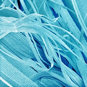
False-colour SEM of retina featuring central fovea
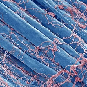
Foveal retina. False-colour scanning electron micrograph of the human retina featuring the central fovea, a crater-like depression in the photosensitive layer of the eye. The foveal retina is the area of greatest visual acuity and contains only cone receptor cells. When an eye looks at an object, that part focused on the fovea is the portion most accurately registered by the brain. This area is particularly rich in blood vessels, which appear root-like, running in, over & outside of the foveal " ridge". Magnification: x24 at 6x7cm size
Science Photo Library features Science and Medical images including photos and illustrations
Media ID 6422336
© PROF. P. MOTTA/DEPT. OF ANATOMY/UNIVERSITY LA SAPIENZA , ROME/SCIENCE PHOTO LIBRARY
Macula Magnified Image Microscopic Photos Retina Sight Subjects Vision Visual Sense False Coloured
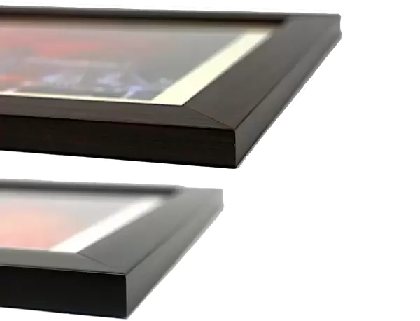
14"x12" (38x32cm) Modern Frame
Bring the wonders of science into your home with Media Storehouse's Framed Prints. This stunning False-colour Scanning Electron Micrograph of the human retina, featuring the central fovea from Science Photo Library, offers a unique and captivating glimpse into the intricacies of the human eye. The central fovea, depicted as a crater-like depression in the photosensitive layer, is the part of the retina responsible for our sharpest vision. This high-quality framed print, with its vibrant and detailed image, is a must-have for any science enthusiast or home decor. Experience the beauty and complexity of the world around us, one frame at a time.
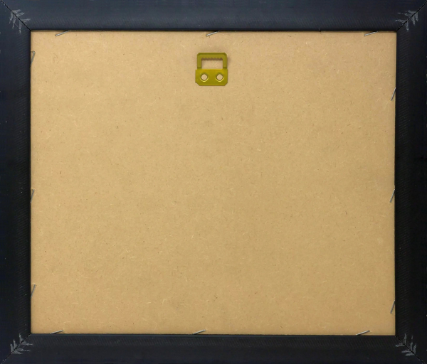
Wood effect frame, card mounted, 10x8 archival quality photo print. Overall outside dimensions 14x12 inches (38x32cm). Environmentally and ozone friendly, 40mm wide x 15mm Polycore® moulding has the look of real wood, is durable and light and easy to hang. Biodegradable and made with non-chlorinated gases (no toxic fumes) it is efficient; producing 100 tons of polystyrene can save 300 tons of trees! Prints are glazed with lightweight, shatterproof, optical clarity acrylic (providing the same general protection from the environment as glass). The back is stapled hardboard with a sawtooth hanger attached. Note: To minimise original artwork cropping, for optimum layout, and to ensure print is secure, the visible print may be marginally smaller
Contemporary Framed and Mounted Prints - Professionally Made and Ready to Hang
Estimated Image Size (if not cropped) is 24.4cm x 18.1cm (9.6" x 7.1")
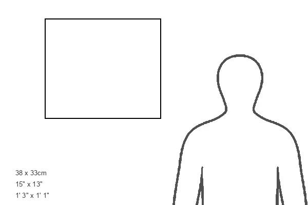
Estimated Product Size is 37.6cm x 32.5cm (14.8" x 12.8")
These are individually made so all sizes are approximate
Artwork printed orientated as per the preview above, with landscape (horizontal) orientation to match the source image.
FEATURES IN THESE COLLECTIONS
> Europe
> Italy
> Lazio
> Rome
EDITORS COMMENTS
This print showcases a false-colour scanning electron micrograph of the human retina, specifically highlighting the central fovea. The foveal retina is an intriguing crater-like depression found in the photosensitive layer of our eyes. It is within this area that we experience our greatest visual acuity, as it exclusively contains cone receptor cells. When we focus our gaze on an object, the part captured by the fovea is meticulously registered by our brain. This region plays a crucial role in providing us with sharp and detailed vision. As we delve into this microscopic image, one cannot help but notice the abundance of root-like blood vessels that intricately weave their way through, over, and outside of the foveal ridge. With a magnification level of x24 at 6x7cm size, this photograph offers us a glimpse into the intricate world of sight and vision. It reminds us how remarkable and complex our visual sense truly is. Furthermore, it highlights key anatomical features such as the macula and fovea which are essential for optimal visual acuity. Through scientific exploration and advancements in imaging technology like SEM (scanning electron microscopy), we gain invaluable insights into subjects like anatomy and physiology. This particular image from Science Photo Library serves as a testament to both its educational value and aesthetic appeal - showcasing nature's wonders hidden within ourselves without any commercial intent behind it.
MADE IN THE UK
Safe Shipping with 30 Day Money Back Guarantee
FREE PERSONALISATION*
We are proud to offer a range of customisation features including Personalised Captions, Color Filters and Picture Zoom Tools
SECURE PAYMENTS
We happily accept a wide range of payment options so you can pay for the things you need in the way that is most convenient for you
* Options may vary by product and licensing agreement. Zoomed Pictures can be adjusted in the Basket.