Fine Art Print : Lenses on brittle star surface, SEM


![]()
Fine Art Prints from Science Photo Library
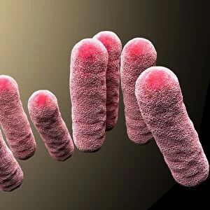
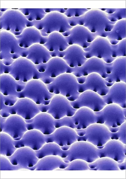
Lenses on brittle star surface, SEM
Brittle star microlenses. Coloured scanning electron micrograph (SEM) of microlenses on the surface of the brittle star Ophiocoma wendtii. These lenses are composed of light-sensitive calcium carbonite crystals. They form a compound eye on the body surface of this marine echinoderm. A network of nerve fibres beneath the lenses detects light signals, allowing the brittle star to detect and escape from predators. The design of the lenses may be used to improve components of optical networks and computer chips. The lenses were discovered by Joanna Aizenberg and her team at Lucent Technologies Bell Labs, USA
Science Photo Library features Science and Medical images including photos and illustrations
Media ID 6466681
© LUCENT TECHNOLOGIES BELL LABS/SCIENCE PHOTO LIBRARY
America Body Brittle Star Compound Eye Crystal Crystals Echinoderm Echinodermata Lens Lenses Light Sensitive Microscopic Optic Optical Optics Purple Sensitivity Surface Us A
A2 (42x59cm) Fine Art Print
Discover the mesmerizing underwater world with our Fine Art Prints from Media Storehouse. This captivating image showcases the intricate beauty of a brittle star, Ophiocoma wendtii, as seen through the lens of a Scanning Electron Microscope from Science Photo Library. Witness the tiny, delicate microlenses that cover the star's surface, each one a testament to nature's intricate design. Bring this stunning piece of marine biology into your home or office and let it inspire awe and wonder. Each print is produced using high-quality materials and techniques to ensure vibrant colors and sharp details, making it a true work of art for your space.
Our Fine Art Prints are printed on 100% acid free, PH neutral paper with archival properties. This printing method is used by museums and art collections to exhibit photographs and art reproductions. Hahnemühle certified studio for digital fine art printing. Printed on 308gsm Photo Rag Paper.
Our fine art prints are high-quality prints made using a paper called Photo Rag. This 100% cotton rag fibre paper is known for its exceptional image sharpness, rich colors, and high level of detail, making it a popular choice for professional photographers and artists. Photo rag paper is our clear recommendation for a fine art paper print. If you can afford to spend more on a higher quality paper, then Photo Rag is our clear recommendation for a fine art paper print.
Estimated Image Size (if not cropped) is 42cm x 53.8cm (16.5" x 21.2")
Estimated Product Size is 42cm x 59.4cm (16.5" x 23.4")
These are individually made so all sizes are approximate
Artwork printed orientated as per the preview above, with portrait (vertical) orientation to match the source image.
EDITORS COMMENTS
This print showcases the intricate beauty of brittle star microlenses, as captured by a coloured scanning electron micrograph (SEM). The surface of the brittle star Ophiocoma wendtii is adorned with these remarkable lenses, which are composed of light-sensitive calcium carbonite crystals. Acting like a compound eye, this network of microlenses enables the marine echinoderm to detect and swiftly evade potential predators. Underneath these delicate lenses lies a complex system of nerve fibers that tirelessly work to interpret incoming light signals. This extraordinary adaptation allows the brittle star to navigate its surroundings with exceptional sensitivity and react promptly when danger arises. Beyond their significance in marine biology, these microlenses hold immense potential for technological advancements. Researchers at Lucent Technologies Bell Labs in the United States, led by Joanna Aizenberg and her team, made this groundbreaking discovery. By studying these natural optical wonders, scientists hope to enhance components within optical networks and computer chips. The vibrant purple hue exhibited in this image adds an element of mystique to an already mesmerizing subject matter. Through this photograph from Science Photo Library, we gain a glimpse into the awe-inspiring world of microscopic optics found within Earth's diverse ecosystem – reminding us once again how nature continues to inspire innovation and push boundaries in science and technology.
MADE IN THE UK
Safe Shipping with 30 Day Money Back Guarantee
FREE PERSONALISATION*
We are proud to offer a range of customisation features including Personalised Captions, Color Filters and Picture Zoom Tools
SECURE PAYMENTS
We happily accept a wide range of payment options so you can pay for the things you need in the way that is most convenient for you
* Options may vary by product and licensing agreement. Zoomed Pictures can be adjusted in the Basket.