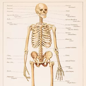
Antique Framed Print : Light micrograph of normal human compact bone


![]()
Framed Photos from Science Photo Library
Light micrograph of normal human compact bone
Light micrograph of a cross-section of normal human compact bone tissue, as found in the shafts of the long bones such as the femur. The rings correspond to concentric bands of calcified material called lamellae. They are centered around an Haversian canal, a channel for the nerves and blood & lymph vessels which runs the entire length of the bone. Each of the small white areas is a lacuna, a hole in the bone which is occupied by an osteocyte or bone cell. The active form of the osteocyte is the osteoblast cell, responsible for bone formation. Each canal and its surrounding lamellae form whats called an Haversian system. Magnification: x200 at 35mm size
Science Photo Library features Science and Medical images including photos and illustrations
Media ID 9303027
© POWER AND SYRED/SCIENCE PHOTO LIBRARY
Bones Compact Bone Haversian Canals Lacuna Osteocyte
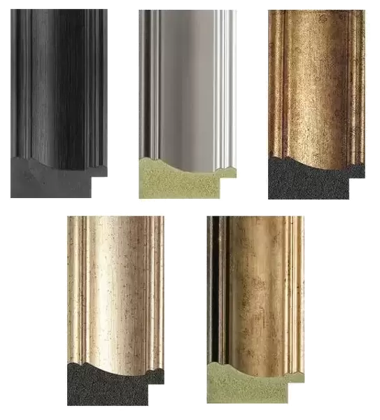
21"x16" (54x41cm) Antique Frame
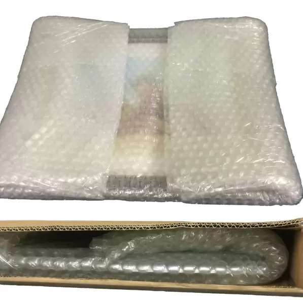
Bevelled wood effect frame, card mounted, 15x10 archival quality photo print. Overall outside dimensions 21x16 inches (54x41cm). Environmentally and ozone friendly, Polycore® moulding has the look of real wood, is durable and light and easy to hang. Biodegradable and made with non-chlorinated gases (no toxic fumes) it is efficient; producing 100 tons of polystyrene can save 300 tons of trees! Prints are glazed with lightweight, shatterproof, optical clarity acrylic (providing the same general protection from the environment as glass). The back is stapled hardboard with a sawtooth hanger attached. Note: To minimise original artwork cropping, for optimum layout, and to ensure print is secure, the visible print may be marginally smaller
Bevelled Wood Effect Framed and Mounted Prints - Professionally Made and Ready to Hang
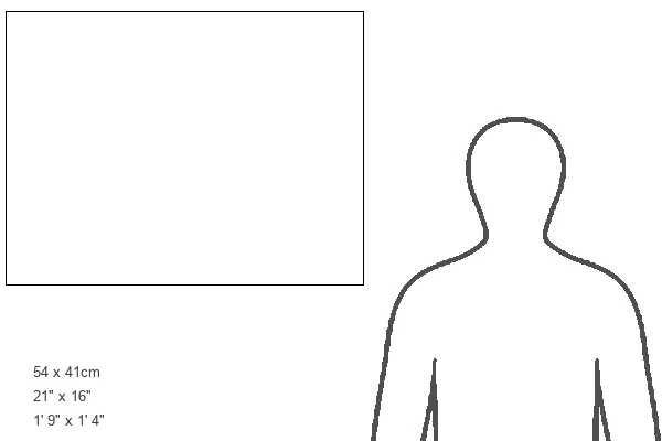
Estimated Image Size (if not cropped) is 37.1cm x 25cm (14.6" x 9.8")
Estimated Product Size is 54cm x 41.4cm (21.3" x 16.3")
These are individually made so all sizes are approximate
Artwork printed orientated as per the preview above, with landscape (horizontal) orientation to match the source image.
EDITORS COMMENTS
This print showcases a mesmerizing view of normal human compact bone tissue. The intricate details captured in this light micrograph provide a glimpse into the complex structure of our bones, specifically those found in the long bones like the femur. The concentric rings visible in the image represent lamellae, which are bands of calcified material. These lamellae encircle an essential component known as the Haversian canal. Serving as a vital channel for nerves and blood & lymph vessels, these canals run throughout the entire length of the bone. Each small white area depicted here is referred to as a lacuna - tiny holes within the bone that house osteocytes or bone cells. Among them, we find active osteoblast cells responsible for bone formation. Together, each Haversian canal and its surrounding lamellae form what is scientifically termed an Haversian system. This remarkable photograph was taken at 200 times magnification using a 35mm size lens by Science Photo Library. Through this visually stunning image, we gain insight into both the beauty and complexity of our skeletal structure. It serves as a reminder of how intricately designed our bodies are and highlights the fascinating world that lies beneath our skin's surface.
MADE IN THE UK
Safe Shipping with 30 Day Money Back Guarantee
FREE PERSONALISATION*
We are proud to offer a range of customisation features including Personalised Captions, Color Filters and Picture Zoom Tools
SECURE PAYMENTS
We happily accept a wide range of payment options so you can pay for the things you need in the way that is most convenient for you
* Options may vary by product and licensing agreement. Zoomed Pictures can be adjusted in the Basket.