
Compact Bone Collection
"Unveiling the Marvels of Compact Bone: A Journey into its Intricate Structure" Delving beneath the surface


All Professionally Made to Order for Quick Shipping
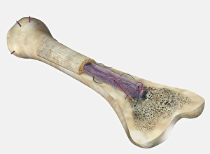
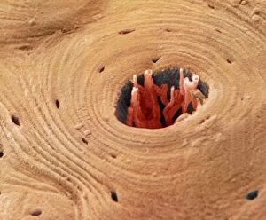
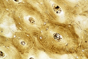
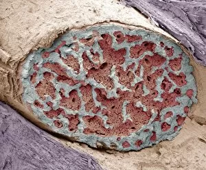
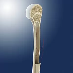
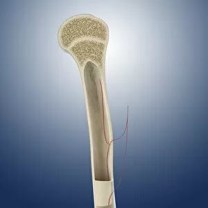
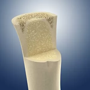
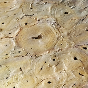
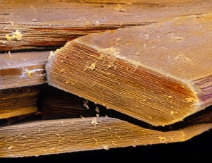
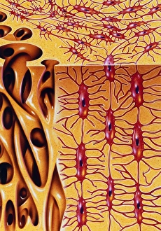
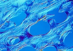
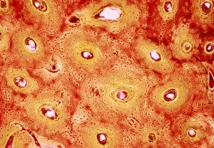
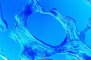
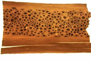
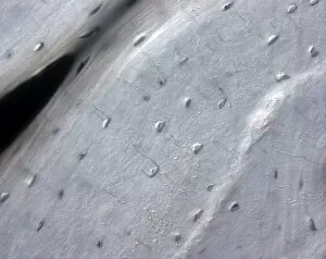
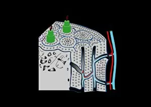
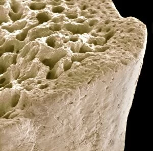
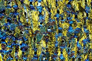
"Unveiling the Marvels of Compact Bone: A Journey into its Intricate Structure" Delving beneath the surface, a light micrograph reveals the intricate beauty of compact bone. This remarkable human bone, as depicted in a cross-section diagram of the Femur, showcases a mesmerizing network of osteons, veins, and marrow. Not limited to humans alone, even whale bone tissue exhibits similar complexity when observed under a light microscope. Its structure mirrors that of our own bones - an awe-inspiring testament to nature's design. A biomedical illustration provides us with further insight into the composition of human bone. The cross-sectional view unveils layers upon layers intricately interwoven with blood vessels and marrow spaces, and is truly a masterpiece in engineering. Intriguingly colored SEM images offer another glimpse into this enigmatic world. These transverse sections highlight the dense arrangement and organization within compact bone like never before seen. Each vibrant hue brings forth an unparalleled visual representation. Light micrographs continue to captivate our imagination as they showcase normal human compact bone in all its glory. The delicate web-like patterns intertwine seamlessly, providing strength and support for our bodies day after day. But not all stories are without their challenges; one such tale lies within a broken finger bone captured by SEM imaging. The fractured edges reveal themselves under scrutiny - reminding us how vital it is to protect these incredible structures that sustain us throughout life's endeavors. Compact bone remains an enduring marvel worth exploring time and again through various lenses - from microscopic views to scientific illustrations. As we unravel its secrets layer by layer, we gain profound appreciation for this essential component that forms the foundation on which we stand tall.