Antique Framed Print > Popular Themes > Human Body
Antique Framed Print : Brain tumour, MRI scan


![]()
Framed Photos from Science Photo Library
Brain tumour, MRI scan
Brain tumour. Coloured orthogonal views (axial left, coronal middle, sagittal right) of a male head, made from magnetic resonance imaging (MRI) scans. The scans reveal a large glioma tumour (yellow-orange), a central nervous system tumour that arises from glial cells. About half of all primary brain tumours are gliomas. MRI uses radio waves and a magnet to obtain " slice" body images
Science Photo Library features Science and Medical images including photos and illustrations
Media ID 6296797
© PASIEKA/SCIENCE PHOTO LIBRARY
Body Central Cerebral Cut Away Diagnosis Diagnostic Dimensional Glioma Imaging Magnetic Nervous Resonance Scan System Brain
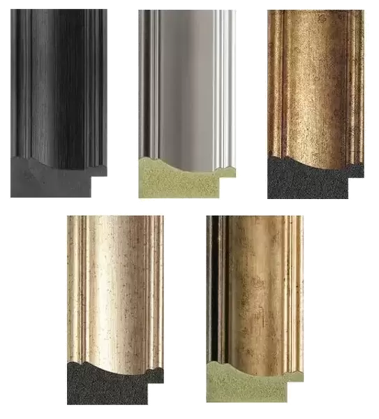
21"x16" (54x41cm) Antique Frame
Bevelled wood effect frame, card mounted, 15x10 archival quality photo print. Overall outside dimensions 21x16 inches (54x41cm). Environmentally and ozone friendly, Polycore® moulding has the look of real wood, is durable and light and easy to hang. Biodegradable and made with non-chlorinated gases (no toxic fumes) it is efficient; producing 100 tons of polystyrene can save 300 tons of trees! Prints are glazed with lightweight, shatterproof, optical clarity acrylic (providing the same general protection from the environment as glass). The back is stapled hardboard with a sawtooth hanger attached. Note: To minimise original artwork cropping, for optimum layout, and to ensure print is secure, the visible print may be marginally smaller
Bevelled Wood Effect Framed and Mounted Prints - Professionally Made and Ready to Hang
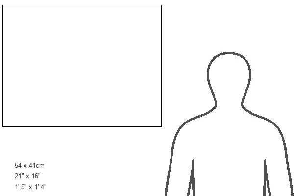
Estimated Image Size (if not cropped) is 37.1cm x 18.4cm (14.6" x 7.2")
Estimated Product Size is 54cm x 41.4cm (21.3" x 16.3")
These are individually made so all sizes are approximate
Artwork printed orientated as per the preview above, with landscape (horizontal) orientation to match the source image.
EDITORS COMMENTS
This print showcases a detailed and colorful representation of a brain tumour, captured through the advanced technology of magnetic resonance imaging (MRI). The image provides an insightful glimpse into the intricate workings of the human central nervous system. The orthogonal views displayed in this print offer different perspectives on the male head, revealing a prominent glioma tumour in vibrant yellow-orange hues. Gliomas are primary brain tumours that originate from glial cells, which play crucial roles in supporting and protecting neurons. It is fascinating to note that approximately half of all primary brain tumours fall under the category of gliomas. This particular MRI scan utilizes radio waves and a powerful magnet to capture "slice" body images with exceptional precision. The three-dimensional nature of this diagnostic tool allows for comprehensive analysis and accurate diagnosis. By peering into this cut-away view, we gain valuable insight into the complex cerebral structures affected by such conditions. Science Photo Library has expertly captured this visually striking image, providing us with both scientific knowledge and artistic appreciation. It serves as a reminder of how medical advancements continue to push boundaries in understanding diseases within our intricate bodies.
MADE IN THE UK
Safe Shipping with 30 Day Money Back Guarantee
FREE PERSONALISATION*
We are proud to offer a range of customisation features including Personalised Captions, Color Filters and Picture Zoom Tools
SECURE PAYMENTS
We happily accept a wide range of payment options so you can pay for the things you need in the way that is most convenient for you
* Options may vary by product and licensing agreement. Zoomed Pictures can be adjusted in the Basket.