Home > Popular Themes > Human Body
Prosthetic hip joint, diagram C016 / 6775

![]()
Wall Art and Photo Gifts from Science Photo Library
Prosthetic hip joint, diagram C016 / 6775
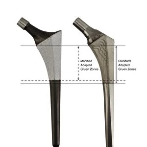
Prosthetic hip joint. Cutaway diagram of a femur (thigh bone) showing a femoral component of a hip prosthesis. This component is implanted in the femur after the head of the femur has been surgically removed. The other components of the hip joint are a rounded ball to fit into the socket implanted in the patients pelvis (not shown). This allows the patient to regain mobility, and is usually done to treat severe osteoarthritis or a broken hip. This is an SHP prosthesis, using bone cement (green). The offset and perimeter parameters are labelled. For this diagram with Gruen zones, see C016/6776
Science Photo Library features Science and Medical images including photos and illustrations
Media ID 9244933
© D & L GRAPHICS / SCIENCE PHOTO LIBRARY
Arthritic Arthritis Arthrology Arthroplasty Artificial Bioceramic Bone Cement Cutaway Device Diagram Femoral Femoral Shaft Femur Hip Implant Hip Replacement Hip Revision Implanted Internal Joint Label Labeled Labelled Labels Metal Offset Orthopaedic Orthopaedics Orthopedic Orthopedics Osteoarthritis Osteological Osteology Perimeter Profile Prostheses Prosthesis Prosthetic Prosthetics Repair Repaired Replacement Shaft Surgery Surgical Total Hip Replacement Treated Treatment Cutouts Section Sectioned
EDITORS COMMENTS
This print showcases a detailed diagram of a prosthetic hip joint, specifically highlighting the femoral component. The cutaway illustration provides an insightful view into the inner workings of this life-changing device. The image depicts a surgically implanted femoral component within the thigh bone (femur), replacing the removed head of the femur. This innovative prosthesis enables patients suffering from severe osteoarthritis or a broken hip to regain mobility and improve their quality of life. Although not shown in this particular diagram, another crucial element of the hip joint is a rounded ball that fits into a socket implanted in the patient's pelvis. Together, these components allow for smooth movement and functionality. Highlighted by its green color, this SHP prosthesis utilizes bone cement technology to ensure stability and durability. The offset and perimeter parameters are clearly labeled, providing valuable information for medical professionals involved in surgical procedures or research. With its intricate details and scientific accuracy, this artwork serves as an invaluable resource for orthopedic specialists, researchers, and students interested in understanding hip replacement surgery and related treatments. Overall, this visually striking print captures both the technological advancements in medicine and the profound impact they have on improving human lives through enhanced mobility and pain relief.
MADE IN THE UK
Safe Shipping with 30 Day Money Back Guarantee
FREE PERSONALISATION*
We are proud to offer a range of customisation features including Personalised Captions, Color Filters and Picture Zoom Tools
SECURE PAYMENTS
We happily accept a wide range of payment options so you can pay for the things you need in the way that is most convenient for you
* Options may vary by product and licensing agreement. Zoomed Pictures can be adjusted in the Basket.