
Arthritic Collection
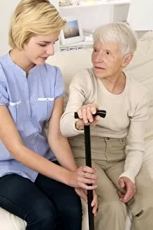
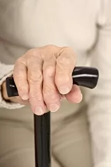
"Living with Arthritis: A Journey of Pain and Hope" Arthritis, a debilitating condition affecting millions worldwide, manifests in various forms throughout the body


All Professionally Made to Order for Quick Shipping
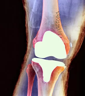
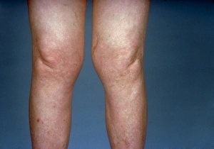
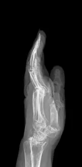
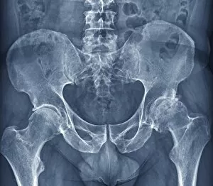
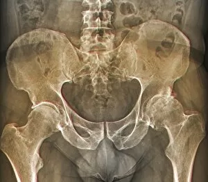
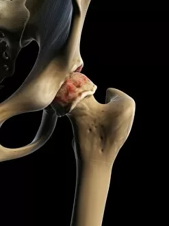
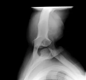
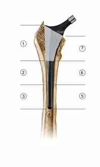
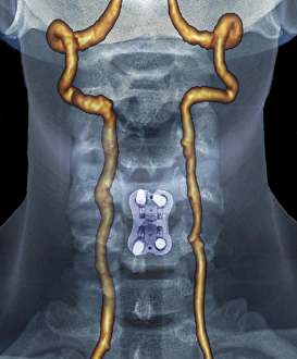
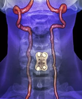
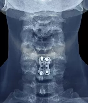
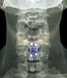
"Living with Arthritis: A Journey of Pain and Hope" Arthritis, a debilitating condition affecting millions worldwide, manifests in various forms throughout the body. From knee joint prosthesis to X-ray images capturing the reality of this ailment, it leaves no part untouched. Doloricure Aninat - French Cure for Rheumatism and Arthritis offers a glimmer of hope amidst the pain. This ancient remedy has provided relief to countless sufferers over centuries. The neck is not spared from arthritis's relentless grip as seen in X-ray C017 / 7389 showcasing arthritis of the neck. The image serves as a reminder that even our most delicate joints are susceptible to this chronic disease. Osteoarthritis takes its toll on knees too; X-ray F006 / 3744 reveals knees affected by this degenerative condition. The view is stark, highlighting the need for interventions such as knee replacements or alternative treatments. Hands bear witness to arthritic struggles as depicted in X-ray C017 / 7171, where an arthritic hand tells its own story of pain and limited mobility. It reminds us that every movement can be an uphill battle for those afflicted. Thoracic spine scoliosis & osteoarthritis further compound the challenges faced by individuals living with arthritis (X-rays C017/0701 & C017/0704). These images serve as visual proof of how multiple conditions can intertwine, amplifying discomfort and impairing daily life. Hip osteoarthritis captures attention through X-rays F006 / 3745 & F006 / 3744; these haunting visuals reveal bones ravaged by time and wear. Yet within these somber frames lies resilience – patients find solace in medical advancements like hip replacements or innovative therapies aimed at restoring function. Artwork becomes a medium through which we explore arthritic knees' emotional impact (artworks F008/2476, F008/2477 & F008/2472).