
Orthopaedic Collection
Orthopaedic care encompasses a wide range of conditions and treatments, all aimed at improving musculoskeletal health


All Professionally Made to Order for Quick Shipping
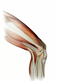
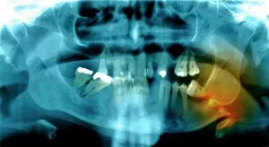
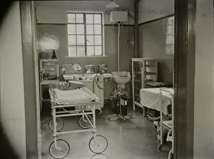
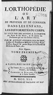
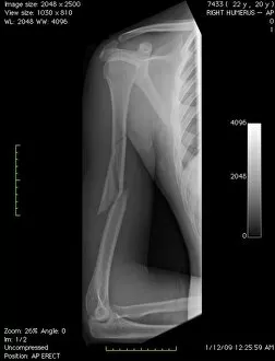
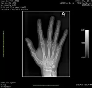
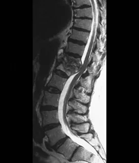
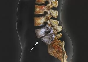
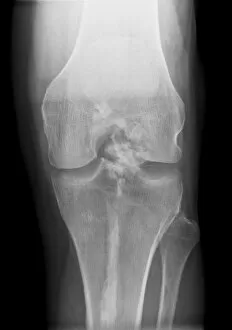
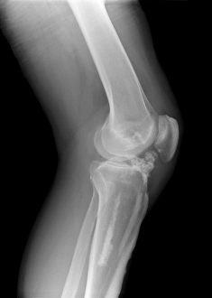
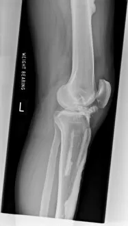
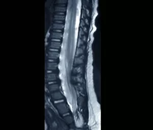
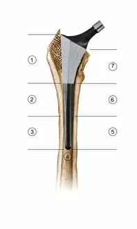
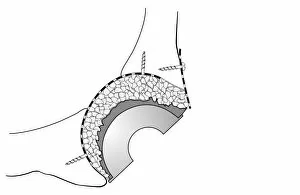
Orthopaedic care encompasses a wide range of conditions and treatments, all aimed at improving musculoskeletal health. From normal knees to damaged ligaments, orthopaedics is dedicated to restoring mobility and relieving pain. In the realm of knee injuries, an X-ray reveals the intricate structure of this joint. Whether it's a normal knee or one with a damaged ligament, orthopaedic interventions can help restore function. An artwork depicting a damaged knee ligament serves as a reminder of the complexity involved in treating such injuries. Shoulder tendinitis is another common ailment that often requires orthopaedic attention. Through an X-ray image, we gain insight into the affected area and understand why proper treatment is crucial for recovery. Headaches can also be linked to orthopaedic issues. A captivating X-ray artwork showcases how these debilitating pains may stem from underlying musculoskeletal problems that require specialized care. The operating theatre symbolizes where skilled surgeons perform intricate procedures to repair fractures like a fractured jawbone shown on an X-ray image. The anaesthetic room adjacent to it highlights the importance of patient comfort during surgeries. Shriners Hospital for Crippled Children in St. Louis Missouri stands as a beacon of hope for children with orthopaedic challenges, offering them comprehensive care and support on their journey towards improved mobility. St Nicholas Homes in Pyrford provides compassionate residential care for individuals requiring long-term orthopaedic assistance. Their dedication ensures patients receive personalized attention tailored to their unique needs. Orthopedic history dates back centuries; even corsets were once used as preventive measures against certain conditions like onanism (masturbation). An illustration from Traite des bandages et appareils by J. B. J Thillaye depicts this historical practice published in 1815 Paris. Even babies are not exempt from orthopedic concerns; Dorothea Lange's poignant photograph captures a baby wearing homemade splints for club feet at an FSA camp in Tulare County, California, 1939.