Home > Science > SEM
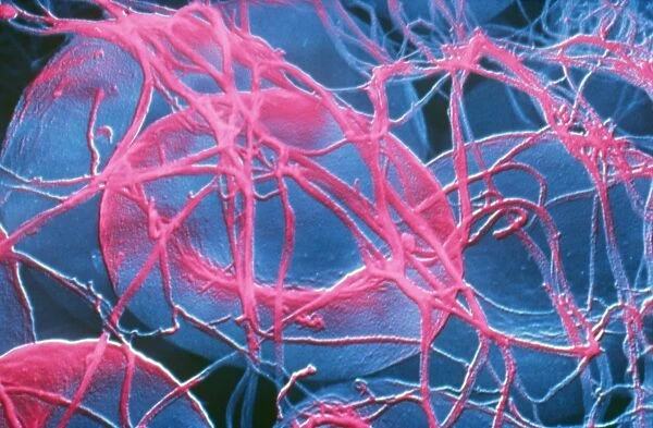
False-colour SEM of a human blood clot
![]()

Wall Art and Photo Gifts from Science Photo Library
False-colour SEM of a human blood clot
False-colour scanning electron micrograph (SEM) of a human blood clot (thrombus), showing strands of fibrin around red blood cells. Blood clots or thrombi form inside blood vessels when there is a defect in the normal haemostatic mechanism. A thrombus composed of red blood cells trapped in a network of fibrin stands is typically formed in areas of stasis (no blood flow) and is known as a coagulation thrombus. Such thrombi are commonly formed in the legs of patients confined to bed in hospital. They may be treated with drugs that interfere with fibrin formation (anticoagulants) or with fibrinolytic drugs that digest fibrin. Mag: x3, 000 at 35mm, x6, 000 at 6x7cm size. RBCs coloured light blue, fibrin pink
Science Photo Library features Science and Medical images including photos and illustrations
Media ID 6421082
© NIBSC/SCIENCE PHOTO LIBRARY
Blood Clotting Coagulation Erythrocyte Fibrin In Blood Clot In Clot Process Red Blood Cell Thrombus False Coloured
FEATURES IN THESE COLLECTIONS
EDITORS COMMENTS
This print showcases a false-colour scanning electron micrograph (SEM) of a human blood clot, providing an intricate view into the complex world within our veins. The image reveals delicate strands of fibrin enveloping vibrant red blood cells, offering a glimpse into the formation and composition of thrombi. Blood clots or thrombi emerge when there is an anomaly in the normal haemostatic mechanism, causing them to form inside blood vessels. In this particular case, we witness a coagulation thrombus composed of red blood cells trapped within a network of fibrin stands. These types of clots typically manifest in areas where blood flow is stagnant, such as the legs of bedridden patients in hospitals. The scientific community has developed various treatments for these potentially harmful formations. Anticoagulants are drugs that interfere with fibrin formation and can be administered to counteract their development. Additionally, fibrinolytic drugs aid in digesting existing fibrin structures. With its magnification at 3,000 times for 35mm prints and 6,000 times for larger 6x7cm prints, this SEM image offers unprecedented detail on the microscopic level. The light blue hue applied to red blood cells contrasts beautifully with the pink shade assigned to fibrin strands—enhancing both aesthetic appeal and scientific clarity. This remarkable photograph from Science Photo Library provides valuable insights into the process behind clotting while highlighting key elements such as erythrocytes (red blood cells), fibrin networks
MADE IN THE UK
Safe Shipping with 30 Day Money Back Guarantee
FREE PERSONALISATION*
We are proud to offer a range of customisation features including Personalised Captions, Color Filters and Picture Zoom Tools
SECURE PAYMENTS
We happily accept a wide range of payment options so you can pay for the things you need in the way that is most convenient for you
* Options may vary by product and licensing agreement. Zoomed Pictures can be adjusted in the Basket.

