Home > Science > SEM
Anthrax bacteria spores

![]()
Wall Art and Photo Gifts from Science Photo Library
Anthrax bacteria spores
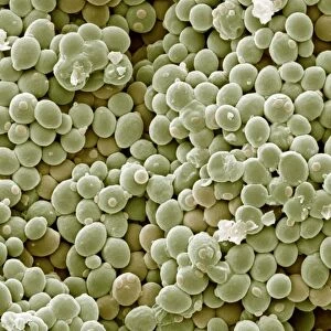
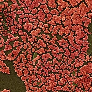
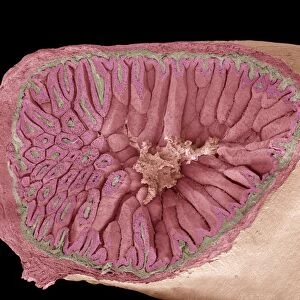
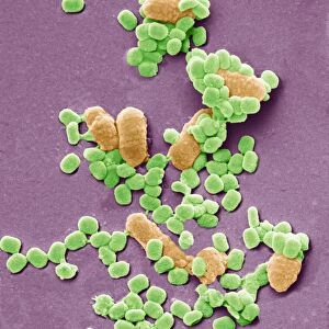
False-colour scanning electron micrograph of spores of the large, rod-shaped bacteria Bacillus anthracis, the causative agent of anthrax in farm animals. The disease is transmitted to man by contact with infected animal hair, hides or excrement. The bacilli attack either the lungs, causing pneumonia (woolsorters disease), or the skin, producing severe ulceration (malignant pustule). Anthrax is treated with antibiotics. In the body the bacteria appear singly or in pairs, but when cultured they link up to form chains, as seen here. Each bacterium produces a single spore, which then germinates into a new organism. Magnification: x2825 at 6x4.5 cm size
Science Photo Library features Science and Medical images including photos and illustrations
Media ID 6293113
© A.B. DOWSETT/SCIENCE PHOTO LIBRARY
Anthrax Bacillus Anthracis Bacteria Bacterial Bacteriology Bacterium Micro Organisms Microbe Microbes Spore Spores Type Micro Biology
EDITORS COMMENTS
This print showcases the intricate world of Anthrax bacteria spores. The false-color scanning electron micrograph reveals the large, rod-shaped Bacillus anthracis bacteria responsible for causing anthrax in farm animals. This disease can be transmitted to humans through contact with infected animal hair, hides, or excrement. The image vividly portrays the two primary ways this menacing bacterium attacks its victims: either by invading the lungs and causing pneumonia (known as woolsorters disease) or by infecting the skin and resulting in severe ulceration known as malignant pustule. Fortunately, antibiotics offer a treatment option against this formidable pathogen. When observed within the human body, these bacilli appear individually or paired up. However, when cultured under laboratory conditions like those captured here, they link together to form chains—a mesmerizing sight that emphasizes their ability to adapt and survive. Each bacterium depicted produces a single spore that eventually germinates into a new organism—an essential part of their life cycle highlighted in this photograph. With an impressive magnification of x2825 at 6x4.5 cm size, we are granted an extraordinary glimpse into the microscopic realm where these microbes thrive. Science Photo Library has once again provided us with a remarkable visual representation of one of nature's most fascinating yet dangerous organisms—Anthrax bacteria spores—which serves as a reminder of both their complexity and potential harm if not properly understood and managed.
MADE IN THE UK
Safe Shipping with 30 Day Money Back Guarantee
FREE PERSONALISATION*
We are proud to offer a range of customisation features including Personalised Captions, Color Filters and Picture Zoom Tools
SECURE PAYMENTS
We happily accept a wide range of payment options so you can pay for the things you need in the way that is most convenient for you
* Options may vary by product and licensing agreement. Zoomed Pictures can be adjusted in the Basket.