Fraud Blocker Skip to main content

Bacterial Collection

"Bacterial: Unveiling the Microscopic World of Disease and Discovery" Step into a time capsule as we journey through history, exploring the captivating realm of bacteria

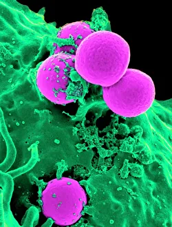
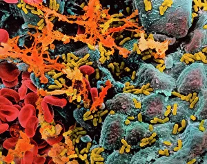
Background imageBacterial Collection: Neutrophil engulfing MRSA, SEM C018 / 8596

Neutrophil engulfing MRSA, SEM C018 / 8596
Neutrophil engulfing MRSA. Coloured scanning electron micrograph (SEM) of a neutrophil white blood cell (green) engulfing methicillin-resistant Staphylococcus aureus bacteria (MRSA, pink)

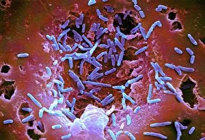
Background imageBacterial Collection: E. coli bacteria, SEM

E. coli bacteria, SEM
E. coli bacteria. Coloured scanning electron micrograph (SEM) of Escherichia coli bacteria (purple) taken from the small intestine of a child. E

Background imageBacterial Collection: Helicobacter pylori bacteria

Helicobacter pylori bacteria
False-colour transmission electron micrograph of the bacterium Helicobacter pylori (formerly called Campylobacter pyloridis)

Background imageBacterial Collection: Plague doctor, 17th century artwork

Plague doctor, 17th century artwork
Plague doctor. 17th century artwork titled Doktor Schnabel von Rom (Beak Doctor from Rome). The text is in Latin. The costumes bronze mask contained aromatic herbs and reduced exposure to bad air

Background imageBacterial Collection: Salmonella bacteria, SEM

Salmonella bacteria, SEM
Salmonella bacteria, coloured scanning electron micrograph (SEM). These Gram-negative rod-shaped bacteria can cause food poisoning when eaten in contaminated food

Background imageBacterial Collection: Calots spinal surgery, 19th century

Calots spinal surgery, 19th century
Calots spinal surgery, 19th-century artwork. This operation is being carried out by the French surgeon Jean-Francois Calot (1861-1944) on a condition known as Potts disease

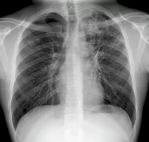
Background imageBacterial Collection: Tuberculosis, X-ray

Tuberculosis, X-ray
Tuberculosis. X-ray of the chest of a 25 year old male patient with pulmonary tuberculosis. Affected areas of the lungs (dark areas) are shown by grainy white patches

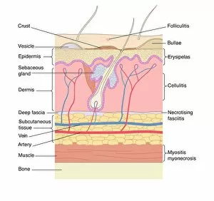
Background imageBacterial Collection: Skin disorders, artwork

Skin disorders, artwork
Skin disorders. Computer artwork showing the structure of the skin, and the layers affected by various skin disorders. Folliculitis is the inflammation of hair follicles; Bullae are blisters;

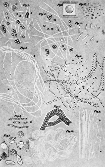
Background imageBacterial Collection: Anthrax cultures, historical diagram

Anthrax cultures, historical diagram
Anthrax cultures. 1876 diagram of Bacillus anthracis (Anthrax) bacteria cultured by Robert Koch. Along with Louis Pasteur, Koch is considered the founder of modern medical bacteriology

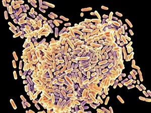
Background imageBacterial Collection: E. coli bacteria, SEM

E. coli bacteria, SEM
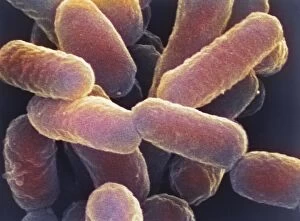
E. coli bacteria. Coloured scanning electron micrograph (SEM) of Escherichia coli bacteria. These bacteria are a normal part of the intestinal flora in humans and other animals

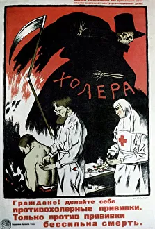
Background imageBacterial Collection: Soviet cholera vaccination poster, 1967

Soviet cholera vaccination poster, 1967
Soviet cholera vaccination poster. Poster which appeared in the USSR in 1967, urging citizens to get themselves vaccinated against cholera

Background imageBacterial Collection: Coloured TEM of Yersinia pestis bacteria

Coloured TEM of Yersinia pestis bacteria
False colour transmission electron micrograph (SEM) of Yersinia pestis, a species of Gram negative, non- motile, capsulated

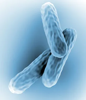
Background imageBacterial Collection: Tuberculosis bacteria

Tuberculosis bacteria. Computer artwork of Mycobacterium tuberculosis bacteria. These Gram- positive rod-shaped bacteria cause the disease tuberculosis

Background imageBacterial Collection: Pseudomonas aeruginosa bacteria, SEM

Pseudomonas aeruginosa bacteria, SEM
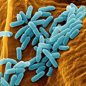
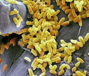
Pseudomonas aeruginosa bacteria, coloured scanning electron micrograph (SEM). These Gram-negative rod-shaped bacteria are found in soil and water, and as normal flora in the human intestine

Background imageBacterial Collection: Spiral spore chain of Streptomyces bacteria

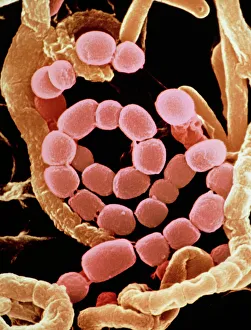
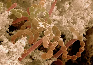
Spiral spore chain of Streptomyces bacteria

Background imageBacterial Collection: Coloured TEM of a Salmonella bacterium

Coloured TEM of a Salmonella bacterium
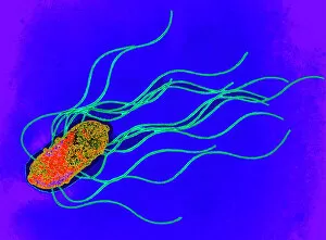
Salmonella sp. Coloured transmission electron micrograph (TEM) of a Salmonella sp. bacterium. The cell is rod-shaped with long hair-like flagellae

Background imageBacterial Collection: Flagellate bacteria

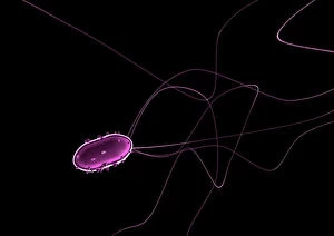
Flagellate bacteria. Computer artwork of a rod- shaped bacteria with polar flagella. Bacteria with a morphology similar to this include Helicobacter pylori and Salmonella sp

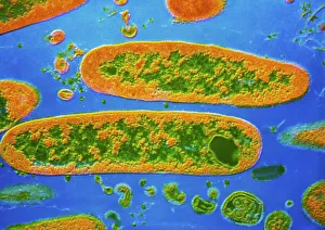
Background imageBacterial Collection: E. coli bacterium

E. coli bacterium
Escherichia coli. Coloured transmission electron micrograph (TEM) of an Escherichia coli bacterium. E. coli are Gram-negative bacilli (rod-shaped) bacteria

Background imageBacterial Collection: E. coli bacteria

E. coli bacteria
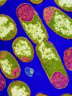
False-colour transmission electron micrograph of the bacterium Escherichia coli, a normal inhabitant of the human intestine

Background imageBacterial Collection: Pseudomonas aeruginosa bacteria, SEM

Pseudomonas aeruginosa bacteria, SEM
Pseudomonas aeruginosa bacteria, coloured scanning electron micrograph (SEM). These Gram-negative rod-shaped bacteria are found in soil and water, and as normal flora in the human intestine

Background imageBacterial Collection: E. coli bacterium, TEM

E. coli bacterium, TEM
E. coli bacterium, coloured transmission electron micrograph (TEM). E. coli are Gram-negative rod-shaped bacteria that are part of the normal flora of the human gut

Background imageBacterial Collection: MRSA resistant Staphylococcus bacteria

MRSA resistant Staphylococcus bacteria
MRSA: resistant Staphylococcus bacteria. Coloured transmission electron micrograph (TEM) of a deadly cluster of MRSA Staphylococcus aureus bacteria

Background imageBacterial Collection: Staphylococcus aureus bacteria

Staphylococcus aureus bacteria
Staphylococcus aureus. Coloured Scanning Electron Micrograph (SEM) of Staphylococcus aureus bacteria. These Gram-positive bacteria cause skin infections

Background imageBacterial Collection: Yoghurt bacteria

Yoghurt bacteria. Coloured scanning electron micrograph (SEM) of the bacteria Streptococcus thermophilus (spherical) and Lactobacillus bulgar- icus (rod-shaped) in live yoghurt. S

Background imageBacterial Collection: Bacterial ribosome

Bacterial ribosome. Computer model showing the secondary structure of a 30S (small) ribosomal sub-unit from the bacteria Thermus thermophilus

Background imageBacterial Collection: Macrophage engulfing TB bacteria, SEM

Macrophage engulfing TB bacteria, SEM
Macrophage engulfing TB bacteria. Coloured scanning electron micrograph (SEM) of a macrophage white blood cell (purple) engulfing a tuberculosis (Mycobacterium tuberculosis) bacterium (pink)

Background imageBacterial Collection: Salmonella bacteria, artwork C013 / 8818

Salmonella bacteria, artwork C013 / 8818
Salmonella bacteria, computer artwork. Salmonella sp. bacteria are gram-negative rod-shaped bacteria that have flagella (hair-like structures) that they use for locomotion

Background imageBacterial Collection: Used dental floss, SEM

Used dental floss, SEM
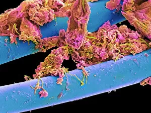
Used dental floss. Coloured scanning electron micrograph (SEM) of dental plaque on strands of used dental floss. Plaque consists of a film of bacteria embedded in a glycoprotein matrix

Background imageBacterial Collection: Neutrophil cell trapping bacteria, SEM

Neutrophil cell trapping bacteria, SEM
Neutrophil cell trapping bacteria. Coloured scanning electron micrograph (SEM) of bacteria (rod-shaped) being trapped by a neutrophil cell

Background imageBacterial Collection: Mycobacterium chelonae bacteria, SEM

Mycobacterium chelonae bacteria, SEM
Mycobacterium chelonae bacteria, coloured scanning electron micrograph (SEM). These Gram-positive rod-shaped bacteria are found in soil and water

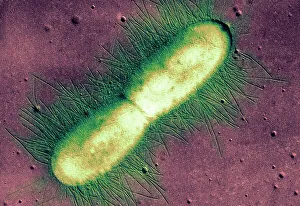
Background imageBacterial Collection: E. coli bacterium, TEM

E. coli bacterium, TEM
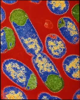
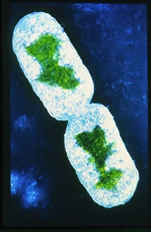
E. coli bacterium. Coloured transmission electron micrograph (TEM) of an Escherichia coli bacterium in the early stages of binary fission, the process by which the bacterium divides

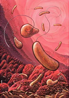
Background imageBacterial Collection: Cholera bacteria, artwork

Cholera bacteria, artwork
Cholera bacteria. Artwork of Vibrio cholerae bacteria in the small intestine. These Gram-negative rod-shaped bacteria have a single polar flagellum (tail-like structure)

Background imageBacterial Collection: Pseudomonas aeruginosa bacteria, SEM

Pseudomonas aeruginosa bacteria, SEM
Pseudomonas aeruginosa bacteria, coloured scanning electron micrograph (SEM). These Gram-negative rod-shaped bacteria are found in soil and water, and as normal flora in the human intestine

Background imageBacterial Collection: Castle Geyser, Yellowstone National Park, Wyoming

Castle Geyser, Yellowstone National Park, Wyoming

Background imageBacterial Collection: Bladder infection

Bladder infection
Bacterial infection of bladder (cystitis). Colour Scanning Electron Micrograph (SEM) of the human bladder showing bacterial infection

Background imageBacterial Collection: Vinegar production, 19th century

Vinegar production, 19th century cutaway artwork. This is a trickling method developed in 1823 by the German chemist Schutzenbach. The barrel is divided into three sections

Background imageBacterial Collection: E. coli 0157: H7 bacteria

E. coli 0157: H7 bacteria
E. coli 0157:H7 bacteria. Coloured scanning electron micrograph (SEM) of Escherichia coli 0157:H7 bacteria, cause of foodborne illness

Background imageBacterial Collection: Salmonella bacterium dividing, SEM

Salmonella bacterium dividing, SEM
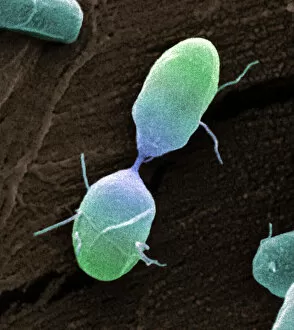
Salmonella bacterium dividing. Coloured scanning electron micrograph (SEM) of of a Salmonella bacterium dividing. The two new daughter cells are seen at upper right and lower left

Background imageBacterial Collection: False-colour TEM of Salmonella typhi

False-colour TEM of Salmonella typhi

Background imageBacterial Collection: False-colour TEM of bacterium E. Coli

False-colour TEM of bacterium E. Coli
False colour transmission electron micrograph of the bacterium Escherichia coli, a normal member of human intestinal flora

Background imageBacterial Collection: E. coli bacterium dividing

E. coli bacterium dividing

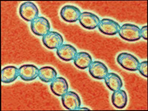
Background imageBacterial Collection: Chains of Streptococcus pyogenes bacteria

Chains of Streptococcus pyogenes bacteria

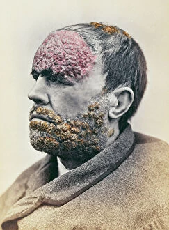
Background imageBacterial Collection: Man suffering from syphilis, from a photograph taken for the Clinique Photographique de

Man suffering from syphilis, from a photograph taken for the Clinique Photographique de
CHT236070 Man suffering from syphilis, from a photograph taken for the Clinique Photographique de l Hopital Saint-Louis, 1868 (colour litho) by French School

Background imageBacterial Collection: Pneumococcal pneumonia. Computer illustration of Streptococcus pneumoniae (pneumococci)

Pneumococcal pneumonia. Computer illustration of Streptococcus pneumoniae (pneumococci) bacteria inside the alveoli of the lungs, causing pneumonia.

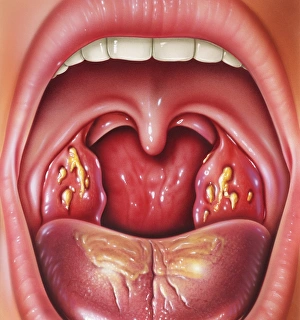
Background imageBacterial Collection: Tonsillitis

Tonsillitis. Artwork of the open mouth and throat of a patient with inflamed tonsils. They are flecked with infected yellow patches, due to tonsillitis

Background imageBacterial Collection: Elevated view of patterns in bacterial mat around Grand Prismatic spring, Midway Geyser Basin

Elevated view of patterns in bacterial mat around Grand Prismatic spring, Midway Geyser Basin, Yellowstone National Park, Wyoming Date: 02-10-2021

Background imageBacterial Collection: Elevated view of Grand Prismatic spring and colorful bacterial mat, Yellowstone National Park

Elevated view of Grand Prismatic spring and colorful bacterial mat, Yellowstone National Park, Wyoming Date: 24-09-2020

Background imageBacterial Collection: Syphilus Bacillus, from a book by Max von Niessen, Leipzig, 1908 (colour litho)

Syphilus Bacillus, from a book by Max von Niessen, Leipzig, 1908 (colour litho)
CHT236063 Syphilus Bacillus, from a book by Max von Niessen, Leipzig, 1908 (colour litho) by German School, (20th century); Bibliotheque Nationale, Paris

Background imageBacterial Collection: Culture of human tuberculosis, from La Tuberculose et son Bacile

Culture of human tuberculosis, from La Tuberculose et son Bacile
CHT235323 Culture of human tuberculosis, from La Tuberculose et son Bacile by Isidore Strauss (1845-96) 1895 (colour litho) by French School

Background imageBacterial Collection: Symptoms of syphilis in men and women, from a medical book, c

Symptoms of syphilis in men and women, from a medical book, c
CHT236068 Symptoms of syphilis in men and women, from a medical book, c.1850-60 (coloured engraving) by French School, (19th century); Bibliotheque Nationale, Paris

Background imageBacterial Collection: Syphilic cluster pustules, from a book by Baron Jean Louis Alibert (1768-1837

Syphilic cluster pustules, from a book by Baron Jean Louis Alibert (1768-1837
CHT236067 Syphilic cluster pustules, from a book by Baron Jean Louis Alibert (1768-1837) 1838 (coloured engraving) by Tresca, Salvadore (1750-1815); Bibliotheque de la Faculte de Medecine, Paris

Background imageBacterial Collection: The facial symptoms of venereal disease, from Traite Complet des Maladies

The facial symptoms of venereal disease, from Traite Complet des Maladies
CHT236066 The facial symptoms of venereal disease, from Traite Complet des Maladies Veneriennes by Philippe Ricord (1800-89) 1851 (colour litho) by French School

Background imageBacterial Collection: Tannerella forsythia bacteria, illustration

Tannerella forsythia bacteria, illustration
Tannerella forsythia bacteria, computer illustration. This anaerobic Gram-negative rod-shaped bacterium was previously called Bacteroides forsythus and Tannerella forsythensis

Background imageBacterial Collection: Dental plaque and tartar

Dental plaque and tartar. Clinical view of a the teeth in a patients lower jaw, with the incisors coated in bacterial plaque and tartar (brown)

Background imageBacterial Collection: Cholera doctor, satirical artwork

Cholera doctor, satirical artwork
Cholera doctor. Satirical artwork of a London doctor gaining wealth (money at lower left) during an 1835 cholera epidemic

Background imageBacterial Collection: Antibiotic action, historical image

Antibiotic action, historical image
Antibiotic action. Historical image taken by the German pathologist Karl Dohle (1855-1928) in 1889, showing the action of an antibiotic compound on Bacillus anthracis (anthrax)

Background imageBacterial Collection: Botulism bacteria

Botulism bacteria. Light micrograph of botulism bacteria (Clostridium botulinum). These bacteria occur naturally in soil. They produce botulinum toxin

Background imageBacterial Collection: RNA-induced silencing complex F006 / 9586

RNA-induced silencing complex F006 / 9586
RNA-induced silencing complex (RISC), molecular model. This complex consists of a bacterial argonaute protein (top) bound to a small interfering RNA (siRNA) molecule (red and blue)

Background imageBacterial Collection: Tuberculosis of the spine, MRI scan

Tuberculosis of the spine, MRI scan
Tuberculosis of the spine. Magnetic resonance imaging (MRI) scan of a sagittal section through the spine of a 74-year-old patient with tuberculosis (TB) of the spine, or Potts disease

Background imageBacterial Collection: Helicobacter pylori bacteria, SEM C016 / 9136

Helicobacter pylori bacteria, SEM C016 / 9136
Helicobacter pylori bacteria. Coloured scanning electron micrograph (SEM) of Helicobacter pylori bacteria (green) in the stomach. H

Background imageBacterial Collection: Helicobacter pylori bacteria, SEM C016 / 9053

Helicobacter pylori bacteria, SEM C016 / 9053
Helicobacter pylori bacteria. Coloured scanning electron micrograph (SEM) of a Helicobacter pylori bacterium (blue) in the stomach. H

Background imageBacterial Collection: Severe gastritis, endoscope view C016 / 3898

Severe gastritis, endoscope view C016 / 3898
Severe gastritis, endoscope view. Gastritis is inflammation of the stomach lining. It causes pain on eating or drinking, and may cause nausea and vomiting

Background imageBacterial Collection: Coloured SEM of Bacillus sp. bacteria

Coloured SEM of Bacillus sp. bacteria
Bacillus sp. Coloured scanning electron micrograph of bacteria cells (Bacillus sp.). These rod- shaped bacteria are found in soil and water and are usually saprotrophic

Background imageBacterial Collection: Acetobacter bacteria, SEM

Acetobacter bacteria, SEM
Vinegar-forming bacteria. Coloured scanning electron micrograph (SEM) of Acetobacter aceti bacteria, which are used in the production of vinegar

Background imageBacterial Collection: Acetobacter and Schizosaccharomyces

Acetobacter and Schizosaccharomyces. Coloured scanning electron micrograph of Acetobacter bacteria and Schizosaccharomyces yeast cells in a culture of Kombucha" tea

Background imageBacterial Collection: Anthrax antibiotics research

Anthrax antibiotics research. Antibiotic drugs (discs) being tested on anthrax bacteria (Bacillus anthracis) colonies being cultured on an agar growth medium in a petri dish

Background imageBacterial Collection: Rod shaped bacillus bacteria

Rod shaped bacillus bacteria
3D-computer artwork of bacillus bacteria. These rod-shaped bacteria (bacilli) are widespread in soil and in the air. Many bacillus bacteria are responsible for food spoilage

Background imageBacterial Collection: Bacteria research

Bacteria research
MODEL RELEASED. Bacteria research. Researcher holding an empty petri dish in front of other dishes containing cultured bacteria (dots)

Background imageBacterial Collection: Chert rock

Chert rock. This carbonaceous chert is from the Barberton greenstone belt in South Africa. The Earths oldest fossil bacteria have been found in this type of rock

Background imageBacterial Collection: Cholera prevention, satirical artwork

Cholera prevention, satirical artwork
Cholera prevention. Satirical artwork portraying a womans efforts to protect herself during an outbreak of cholera. Cholera is a bacterial infection of the intestines that was often fatal before

Background imageBacterial Collection: Bacterial cell structure, artwork

Bacterial cell structure, artwork
Bacterial cell structure. Computer artwork showing the cell structure and components (organelles) of a typical rod-shaped bacteria (bacillus). Not all bacteria have a flagellum (long, tail-like)

Background imageBacterial Collection: Bacillus subtilis bacteria, SEM

Bacillus subtilis bacteria, SEM
Bacillus subtilis. Coloured scanning electron micrograph (SEM) of Bacillus subtilis bacteria. B. subtilis, or hay bacillus, is an aerobic, gram-positive bacillus(rod-shaped bacterium)

Background imageBacterial Collection: Lactobacillus bacteria, SEM

Lactobacillus bacteria, SEM
Lactobacillus sp. bacteria, coloured scanning electron micrograph (SEM). Lactobacillus bacteria are probiotic bacteria, which are useful for human and animal health as they produce bacteriocins

Background imageBacterial Collection: Clostridium perfringens bacterium with spore

Clostridium perfringens bacterium with spore
False-colour transmission electron micrograph (TEM) of a Clostridium perfringens bacterium with endospore. This species of Gram-positive

Background imageBacterial Collection: Bacteria on lung hairs

Bacteria on lung hairs
Bacteria in a lung. Computer artwork of bacteria (yellow) on cilia (hair-like structures) in a lung. Bacterial infection of the lungs can cause potentially fatal pneumonia

Background imageBacterial Collection: Proteus mirabilis bacterium

Proteus mirabilis bacterium
False-colour transmission electron micrograph of the bacterium Proteus mirabilis. It is an enterobacterium which is present normally in the human intestine

Background imageBacterial Collection: Spirochete bacteria, TEM

Spirochete bacteria, TEM
Spirochete bacteria, coloured transmission electron micrograph (TEM). Spirochetes are bacteria with helically coiled cells

Background imageBacterial Collection: Coloured SEM of Clostridium botulinum

Coloured SEM of Clostridium botulinum
Food poisoning bacterium. Coloured scanning electron micrograph of the Gram-positive anaerobic bacteria Clostridium botulinum, responsible for serious food poisoning in humans. C

Background imageBacterial Collection: Rod-shaped bacteria

Rod-shaped bacteria. Computer artwork of rod- shaped bacteria (bacilli). Typical bacilli are E. coli and Salmonella bacteria, though there are many others as well

Background imageBacterial Collection: Nitrobacter sp bacteria, TEM

Nitrobacter sp bacteria, TEM

Background imageBacterial Collection: Plague bacteria

Plague bacteria. Light micrograph of Gram- stained Yersinia pestis bacteria. This bacterium is the cause of bubonic plague (generally agreed to have been the Black Death of the Middle Ages)

Background imageBacterial Collection: Mammoth Hot Springs Terraces in the Early Morning, Mammoth Hot Springs, Yellowstone National Park

Mammoth Hot Springs Terraces in the Early Morning, Mammoth Hot Springs, Yellowstone National Park, Wyoming, USA

Background imageBacterial Collection: Grand Prismatic Spring (3), Middle Geyser Basin, Yellowstone National Park, Wyoming, USA

Grand Prismatic Spring (3), Middle Geyser Basin, Yellowstone National Park, Wyoming, USA


All Professionally Made to Order for Quick Shipping

"Bacterial: Unveiling the Microscopic World of Disease and Discovery" Step into a time capsule as we journey through history, exploring the captivating realm of bacteria. From the haunting presence of plague doctors in 17th-century artwork to cutting-edge scientific images captured by scanning electron microscopes (SEM), prepare to be fascinated by these tiny yet formidable organisms. Witness the battle between our immune system and Methicillin-resistant Staphylococcus aureus (MRSA) as a brave neutrophil engulfs it under SEM C018/8596. Marvel at the groundbreaking Calots spinal surgery from the 19th century, which revolutionized medical practices despite lurking bacterial threats. Peering through an X-ray, tuberculosis reveals its sinister grip on human lungs while skin disorders come alive in vivid artwork, reminding us of bacteria's impact on our bodies. E. coli bacteria, magnified under SEM, showcases their intricate structures that belie their harmful potential. Travel back to Soviet Russia in 1967 with a cholera vaccination poster that symbolizes humanity's fight against bacterial epidemics. Observe salmonella bacteria under SEM; their deceptively beautiful appearance belies their ability to cause severe illness. Delve into historical diagrams depicting anthrax cultures and learn about Yersinia pestis - responsible for devastating pandemics throughout history. The tuberculosis bacterium reminds us of its long-standing presence as one of humanity's greatest adversaries. Finally, witness nature's artistic flair with spiral spore chains formed by Streptomyces bacteria – showcasing both beauty and resilience within this microscopic world. Through these glimpses into bacterial realms past and present, we gain insight into our ongoing struggle against infectious diseases. Let this exploration ignite curiosity about microbial lifeforms that shape our existence – forever reminding us how knowledge can empower us in combating these invisible foes.

© Copyright Media Storehouse, All Rights Reserved 2003 - 2024
Facebook     Pinterest     Card Payments     Reviews IO