
Premium Framed Print : Treatment using X-rays, 1897


![]()
Framed Photos from Science Photo Library
Treatment using X-rays, 1897
Treatment using X-rays. 19th-century artwork of a patient in bed being treated by X-rays from the machine at right. At the time (1897), X-rays had been systematically studied since 1895 by Wilhelm Roentgen, but it was not yet realised that excessive exposure is dangerous. This machine was constructed in Paris by French instrument makers Eugene Ducretet (1844-1915) and L. Lejeune. Artwork from the first volume (1897) of La Revue Scientifique et Industrielle by French chemist and inventor Jules-Louis Breton (1872-1940)
Science Photo Library features Science and Medical images including photos and illustrations
Media ID 9210661
© SCIENCE PHOTO LIBRARY
1897 Apparatus Carcinogenic Clinic Danger Dangerous Device France French Hazard Hospital Jules Louis Breton La Revue Scientifique Et Industrielle Patient Risk Scientific And Industrial Review Treating Treatment X Ray Machine Lejeune Physical Radiotherapy
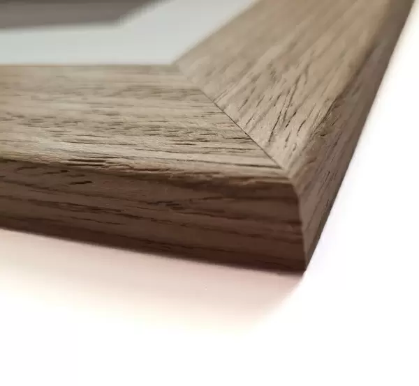
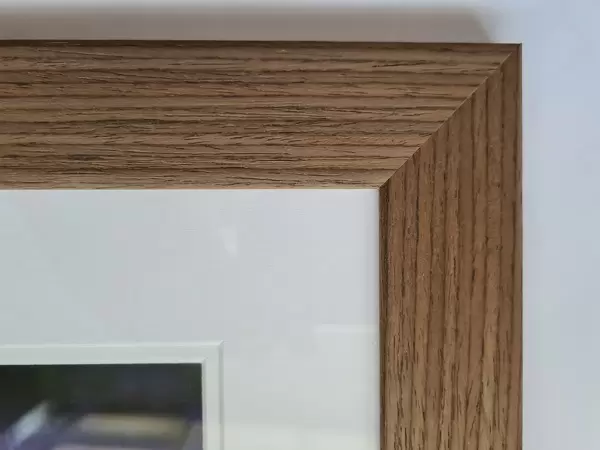
17"x15" (43x38cm) Premium Frame
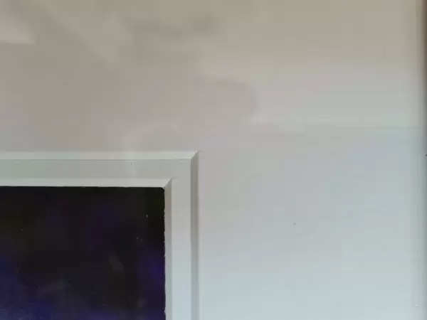
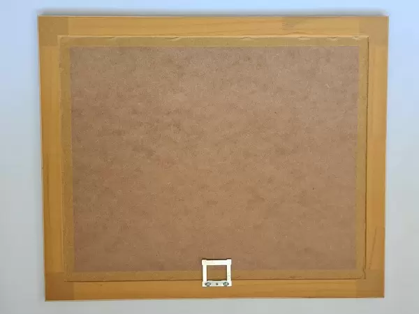
FSC real wood frame with double mounted 10x8 print. Double mounted with white conservation mountboard. Frame moulding comprises stained composite natural wood veneers (Finger Jointed Pine) 39mm wide by 21mm thick. Archival quality Fujifilm CA photo paper mounted onto 1mm card. Overall outside dimensions are 17x15 inches (431x381mm). Rear features Framing tape to cover staples, 50mm Hanger plate, cork bumpers. Glazed with durable thick 2mm Acrylic to provide a virtually unbreakable glass-like finish. Acrylic Glass is far safer, more flexible and much lighter than typical mineral glass. Moreover, its higher translucency makes it a perfect carrier for photo prints. Acrylic allows a little more light to penetrate the surface than conventional glass and absorbs UV rays so that the image and the picture quality doesn't suffer under direct sunlight even after many years. Easily cleaned with a damp cloth. Please note that, to prevent the paper falling through the mount window and to prevent cropping of the original artwork, the visible print may be slightly smaller to allow the paper to be securely attached to the mount without any white edging showing and to match the aspect ratio of the original artwork.
FSC Real Wood Frame and Double Mounted with White Conservation Mountboard - Professionally Made and Ready to Hang
Estimated Image Size (if not cropped) is 24.4cm x 21.7cm (9.6" x 8.5")
Estimated Product Size is 43.1cm x 38.1cm (17" x 15")
These are individually made so all sizes are approximate
Artwork printed orientated as per the preview above, with landscape (horizontal) orientation to match the source image.
EDITORS COMMENTS
This 19th-century artwork captures a pivotal moment in medical history - the early use of X-rays for treatment. The scene depicts a patient lying in bed, undergoing a revolutionary procedure using X-rays emitted from the machine on the right. Created in 1897 by French chemist and inventor Jules-Louis Breton, this illustration showcases an era when X-rays were still being explored and their potential dangers were not yet fully understood. At that time, Wilhelm Roentgen had been diligently studying X-rays since 1895, but it was not widely recognized that excessive exposure could be hazardous. The intricate machine used in this treatment was constructed by renowned French instrument makers Eugene Ducretet and L. Lejeune, reflecting the technological advancements of the period. The historical significance of this artwork lies in its portrayal of cutting-edge medical technology during the late 1800s. It serves as a reminder of how far we have come in understanding radiation's risks and benefits for patients' well-being. Today, we recognize that proper precautions must be taken to ensure safe usage of X-ray machines. This image offers us a glimpse into our past while highlighting the importance of ongoing scientific research and development within medicine.
MADE IN THE UK
Safe Shipping with 30 Day Money Back Guarantee
FREE PERSONALISATION*
We are proud to offer a range of customisation features including Personalised Captions, Color Filters and Picture Zoom Tools
FREE COLORIZATION SERVICE
You can choose advanced AI Colorization for this picture at no extra charge!
SECURE PAYMENTS
We happily accept a wide range of payment options so you can pay for the things you need in the way that is most convenient for you
* Options may vary by product and licensing agreement. Zoomed Pictures can be adjusted in the Basket.