Premium Framed Print : Muscles of the foot


![]()
Framed Photos from Science Photo Library
Muscles of the foot
Muscles of the foot, historical artwork. The figure at top left shows the first layer of muscles (red) in the sole of the foot. The skin and fascia (connective tissue) have been removed. At top right is the second layer, and at bottom right is the third and deepest layer of muscles in the sole of the foot. The underside of the heel bone (os calcis) is at top. Flexor and abductor muscles that move the toes attach to the heel bone. The figure at bottom left shows the muscles on the upper surface of the foot, which include the extensor muscles of the toes. Published in The Muscles of the Human Body... by Jones Quain in 1836
Science Photo Library features Science and Medical images including photos and illustrations
Media ID 6448613
© SHEILA TERRY/SCIENCE PHOTO LIBRARY
1836 19th Abductor Bones Carpal Tunnel Syndrome Dorsal Extensor Feet Flexor Foot Heel Historical Image Imagery Layer Layers Ligament Ligaments Muscle System Muscles Muscular Nineteenth Century Plane Planes Sole Tendon Tendons Toes Under Side Comments Jones Musculature Quain
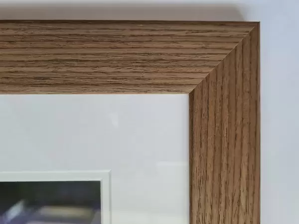
23"x19" (58x48cm) Premium Frame
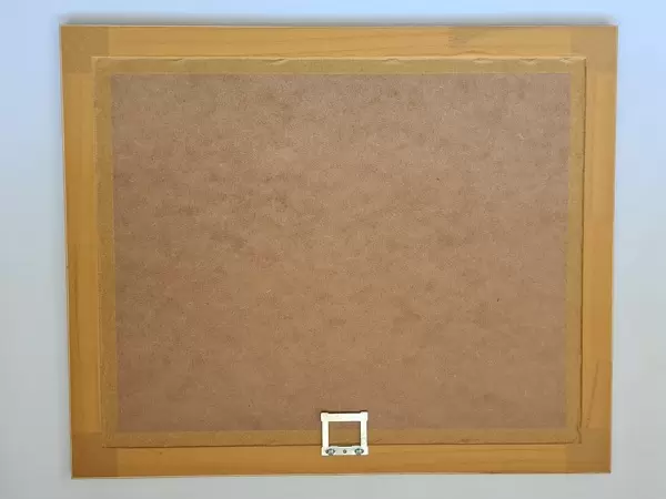
FSC real wood frame with double mounted 16x12 print. Double mounted with white conservation mountboard. Frame moulding comprises stained composite natural wood veneers (Finger Jointed Pine) 39mm wide by 21mm thick. Archival quality Fujifilm CA photo paper mounted onto 1mm card. Overall outside dimensions are 23x19 inches (584x482mm). Rear features Framing tape to cover staples, 50mm Hanger plate, cork bumpers. Glazed with durable thick 2mm Acrylic to provide a virtually unbreakable glass-like finish. Acrylic Glass is far safer, more flexible and much lighter than typical mineral glass. Moreover, its higher translucency makes it a perfect carrier for photo prints. Acrylic allows a little more light to penetrate the surface than conventional glass and absorbs UV rays so that the image and the picture quality doesn't suffer under direct sunlight even after many years. Easily cleaned with a damp cloth. Please note that, to prevent the paper falling through the mount window and to prevent cropping of the original artwork, the visible print may be slightly smaller to allow the paper to be securely attached to the mount without any white edging showing and to match the aspect ratio of the original artwork.
FSC Real Wood Frame and Double Mounted with White Conservation Mountboard - Professionally Made and Ready to Hang
Estimated Image Size (if not cropped) is 25.8cm x 39.6cm (10.2" x 15.6")
Estimated Product Size is 48.2cm x 58.4cm (19" x 23")
These are individually made so all sizes are approximate
Artwork printed orientated as per the preview above, with portrait (vertical) orientation to match the source image.
EDITORS COMMENTS
This historical artwork, titled "Muscles of the Foot" provides a detailed and intricate illustration of the muscular structure within our feet. Created by Jones Quain in 1836, this 19th-century print showcases three layers of muscles found in the sole of the foot. In the top left corner, we observe the first layer (depicted in red) after removing the skin and fascia. Moving to the top right, we encounter the second layer, followed by the third and deepest layer at bottom right. The underside of the heel bone (os calcis) is prominently displayed at the top, where flexor and abductor muscles responsible for toe movement attach. The figure at bottom left shifts our focus to explore muscles on the upper surface of our feet. This includes extensor muscles that play a crucial role in toe extension. With its meticulous attention to detail, this artwork offers valuable insights into foot anatomy from an earlier era. Published as part of "The Muscles of Human Body" series by Jones Quain, this image serves as a remarkable reference for anatomical study even today. It portrays various planes, tendons, ligaments while providing physiological references. With its rich imagery and comprehensive depiction of foot musculature dating back centuries ago, this historical image continues to be a valuable resource for those interested in understanding human anatomy or exploring medical history's artistic representations.
MADE IN THE UK
Safe Shipping with 30 Day Money Back Guarantee
FREE PERSONALISATION*
We are proud to offer a range of customisation features including Personalised Captions, Color Filters and Picture Zoom Tools
SECURE PAYMENTS
We happily accept a wide range of payment options so you can pay for the things you need in the way that is most convenient for you
* Options may vary by product and licensing agreement. Zoomed Pictures can be adjusted in the Basket.