Premium Framed Print > Animals > Insects > Butterflies > Bagworm
Premium Framed Print : Light micrograph of the blood fluke Schistosoma


![]()
Framed Photos from Science Photo Library
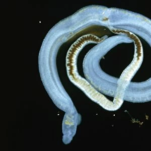
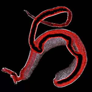
Light micrograph of the blood fluke Schistosoma
Light micrograph of adult intestinal blood flukes, Schistosoma mansoni, cause of schistosomasis, commonly known as bilharzia. The adults (male thick & bluish, female white threadlike) normally live in pairs in blood vessels of the small intestine, causing dysentery & diarrhoea. Their spiked eggs cause anaemia, inflammation & tissue scarring. The larvae develop in freshwater snails (intermediate host) & are released into the water. Humans (final host) are infected while bathing or working in contaminated water. The dark brown coloration, particularly noticable in the female, is half digested blood from a previous meal. Magnification: x5 at 35mm size. These specimens were taken from the liver of a
Science Photo Library features Science and Medical images including photos and illustrations
Media ID 6468663
© SINCLAIR STAMMERS/SCIENCE PHOTO LIBRARY
Blood Flat Worm Fluke Parasite Parasitic Platyhelminthes Schistosoma Mansoni
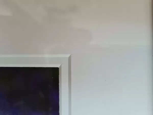
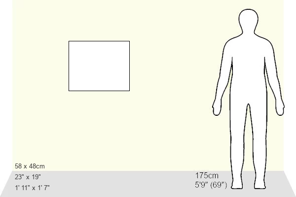
23"x19" (58x48cm) Premium Frame
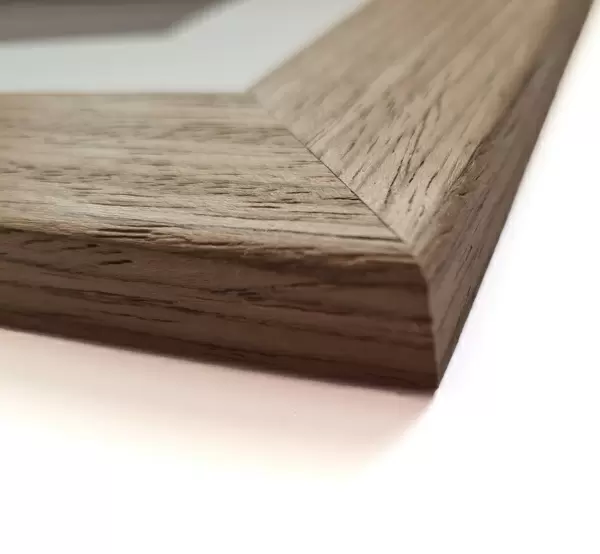
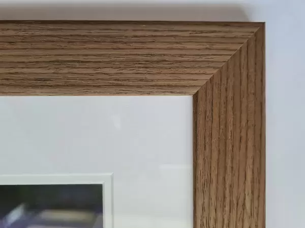
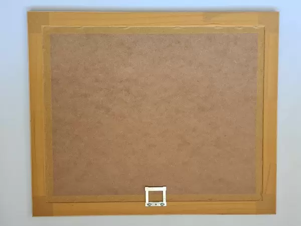
FSC real wood frame with double mounted 16x12 print. Double mounted with white conservation mountboard. Frame moulding comprises stained composite natural wood veneers (Finger Jointed Pine) 39mm wide by 21mm thick. Archival quality Fujifilm CA photo paper mounted onto 1mm card. Overall outside dimensions are 23x19 inches (584x482mm). Rear features Framing tape to cover staples, 50mm Hanger plate, cork bumpers. Glazed with durable thick 2mm Acrylic to provide a virtually unbreakable glass-like finish. Acrylic Glass is far safer, more flexible and much lighter than typical mineral glass. Moreover, its higher translucency makes it a perfect carrier for photo prints. Acrylic allows a little more light to penetrate the surface than conventional glass and absorbs UV rays so that the image and the picture quality doesn't suffer under direct sunlight even after many years. Easily cleaned with a damp cloth. Please note that, to prevent the paper falling through the mount window and to prevent cropping of the original artwork, the visible print may be slightly smaller to allow the paper to be securely attached to the mount without any white edging showing and to match the aspect ratio of the original artwork.
FSC Real Wood Frame and Double Mounted with White Conservation Mountboard - Professionally Made and Ready to Hang
Estimated Image Size (if not cropped) is 39.6cm x 26.7cm (15.6" x 10.5")
Estimated Product Size is 58.4cm x 48.2cm (23" x 19")
These are individually made so all sizes are approximate
Artwork printed orientated as per the preview above, with landscape (horizontal) orientation to match the source image.
EDITORS COMMENTS
This print showcases a light micrograph of the blood fluke Schistosoma, shedding light on the fascinating world of parasitic organisms. The image reveals adult intestinal blood flukes, specifically Schistosoma mansoni, which are notorious for causing schistosomiasis, commonly known as bilharzia. The male flukes appear thick and bluish in color, while their female counterparts take on a delicate white threadlike form. Typically found living in pairs within the blood vessels of the small intestine, these parasites wreak havoc by inducing dysentery and diarrhea among their unfortunate hosts. Moreover, their spiked eggs contribute to anemia, inflammation, and tissue scarring. The life cycle of these intriguing creatures involves developing within freshwater snails before being released into water as larvae. Humans become infected when they come into contact with contaminated water during activities such as bathing or working. Notably visible in this image is the dark brown coloration present in the female fluke's body - evidence of half-digested blood from a previous meal. With a magnification factor of x5 at 35mm size, this photograph offers an up-close look at these remarkable specimens extracted from a liver sample. Captured by Science Photo Library's expert lensmen specializing in nature photography and zoology subjects like wild animals and invertebrates such as platyhelminthes (flatworms), this print provides valuable insight into the intricate world of parasitic organisms without any commercial intent behind it.
MADE IN THE UK
Safe Shipping with 30 Day Money Back Guarantee
FREE PERSONALISATION*
We are proud to offer a range of customisation features including Personalised Captions, Color Filters and Picture Zoom Tools
SECURE PAYMENTS
We happily accept a wide range of payment options so you can pay for the things you need in the way that is most convenient for you
* Options may vary by product and licensing agreement. Zoomed Pictures can be adjusted in the Basket.