Fine Art Print > Animals > Insects > Butterflies > Bagworm
Fine Art Print : Light micrograph of the blood fluke Schistosoma


![]()
Fine Art Prints from Science Photo Library
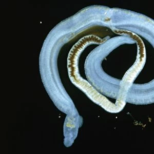
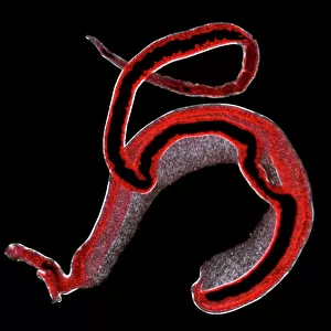
Light micrograph of the blood fluke Schistosoma
Light micrograph of adult intestinal blood flukes, Schistosoma mansoni, cause of schistosomasis, commonly known as bilharzia. The adults (male thick & bluish, female white threadlike) normally live in pairs in blood vessels of the small intestine, causing dysentery & diarrhoea. Their spiked eggs cause anaemia, inflammation & tissue scarring. The larvae develop in freshwater snails (intermediate host) & are released into the water. Humans (final host) are infected while bathing or working in contaminated water. The dark brown coloration, particularly noticable in the female, is half digested blood from a previous meal. Magnification: x5 at 35mm size. These specimens were taken from the liver of a
Science Photo Library features Science and Medical images including photos and illustrations
Media ID 6468663
© SINCLAIR STAMMERS/SCIENCE PHOTO LIBRARY
Blood Flat Worm Fluke Parasite Parasitic Platyhelminthes Schistosoma Mansoni
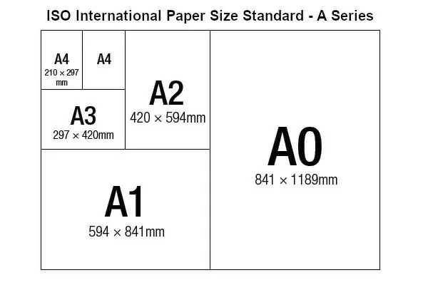
A2 (42x59cm) Fine Art Print
Discover the intricate beauty of the natural world with Media Storehouse's Fine Art Prints. This captivating image showcases a light micrograph of the adult intestinal blood flukes, Schistosoma mansoni, from Science Photo Library. Known for causing the parasitic disease schistosomiasis or bilharzia, these organisms reveal a mesmerizing complexity when viewed under a microscope. Our Fine Art Prints are meticulously printed on premium quality paper, ensuring vibrant colors and stunning detail. Bring the wonders of science into your home or office and ignite your imagination with this unique and thought-provoking piece.
Our Fine Art Prints are printed on 100% acid free, PH neutral paper with archival properties. This printing method is used by museums and art collections to exhibit photographs and art reproductions. Hahnemühle certified studio for digital fine art printing. Printed on 308gsm Photo Rag Paper.
Our fine art prints are high-quality prints made using a paper called Photo Rag. This 100% cotton rag fibre paper is known for its exceptional image sharpness, rich colors, and high level of detail, making it a popular choice for professional photographers and artists. Photo rag paper is our clear recommendation for a fine art paper print. If you can afford to spend more on a higher quality paper, then Photo Rag is our clear recommendation for a fine art paper print.
Estimated Image Size (if not cropped) is 59.4cm x 40cm (23.4" x 15.7")
Estimated Product Size is 59.4cm x 42cm (23.4" x 16.5")
These are individually made so all sizes are approximate
Artwork printed orientated as per the preview above, with landscape (horizontal) orientation to match the source image.
EDITORS COMMENTS
This print showcases a light micrograph of the blood fluke Schistosoma, shedding light on the fascinating world of parasitic organisms. The image reveals adult intestinal blood flukes, specifically Schistosoma mansoni, which are notorious for causing schistosomiasis, commonly known as bilharzia. The male flukes appear thick and bluish in color, while their female counterparts take on a delicate white threadlike form. Typically found living in pairs within the blood vessels of the small intestine, these parasites wreak havoc by inducing dysentery and diarrhea among their unfortunate hosts. Moreover, their spiked eggs contribute to anemia, inflammation, and tissue scarring. The life cycle of these intriguing creatures involves developing within freshwater snails before being released into water as larvae. Humans become infected when they come into contact with contaminated water during activities such as bathing or working. Notably visible in this image is the dark brown coloration present in the female fluke's body - evidence of half-digested blood from a previous meal. With a magnification factor of x5 at 35mm size, this photograph offers an up-close look at these remarkable specimens extracted from a liver sample. Captured by Science Photo Library's expert lensmen specializing in nature photography and zoology subjects like wild animals and invertebrates such as platyhelminthes (flatworms), this print provides valuable insight into the intricate world of parasitic organisms without any commercial intent behind it.
MADE IN THE UK
Safe Shipping with 30 Day Money Back Guarantee
FREE PERSONALISATION*
We are proud to offer a range of customisation features including Personalised Captions, Color Filters and Picture Zoom Tools
SECURE PAYMENTS
We happily accept a wide range of payment options so you can pay for the things you need in the way that is most convenient for you
* Options may vary by product and licensing agreement. Zoomed Pictures can be adjusted in the Basket.