
Premium Framed Print : Knee bones and ligaments


![]()
Framed Photos from Science Photo Library
Knee bones and ligaments
Knee bones and ligaments. Historical anatomical artwork of knee bones (yellow) and ligaments (pale blue). Ligaments are bands of fibrous tissue that hold bones together at joints. An undissected knee is seen from four sides: front (upper left); back (upper right); outer side (centre left); and inner side (centre right). Ligaments obscure the kneecap (patella). The knee (frontal view) is increasingly dissected by removing the synovial membrane (upper centre) and the patella (lower left). The floor of the knee joint (from above) is at lower centre. A vertical section through the knee is at lower right. Artwork from The Bones and Ligaments of the Human Body (Ed. Jones Quain, London, 1842)
Science Photo Library features Science and Medical images including photos and illustrations
Media ID 6419452
© SHEILA TERRY/SCIENCE PHOTO LIBRARY
1842 Anterior Arthrological Arthrology Behind Bones Book Connective Tissue Dissected Dissection Drawing External Front Frontal Internal Joint Joints Jones Quain Knee Knee Cap Lateral Ligament Ligaments Patella Posterior Profile Rear Side Skeletal Synovial Membrane Text Book Section Sectioned
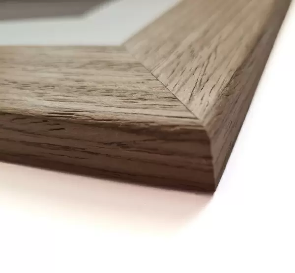
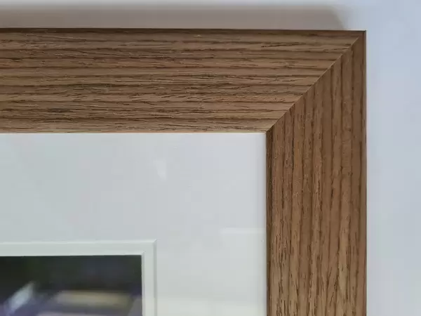
23"x19" (58x48cm) Premium Frame
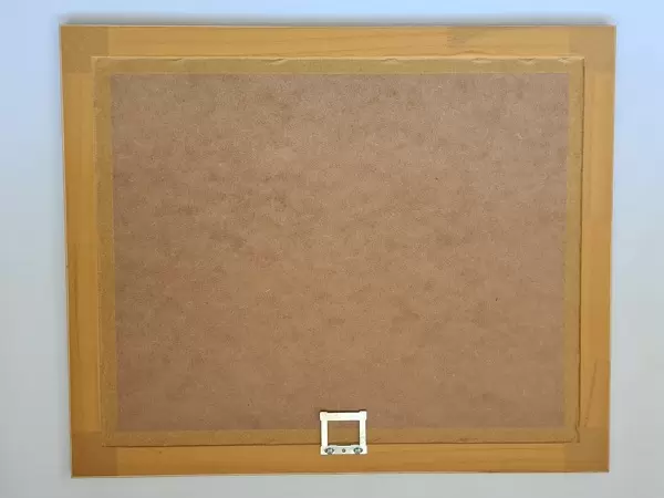
FSC real wood frame with double mounted 16x12 print. Double mounted with white conservation mountboard. Frame moulding comprises stained composite natural wood veneers (Finger Jointed Pine) 39mm wide by 21mm thick. Archival quality Fujifilm CA photo paper mounted onto 1mm card. Overall outside dimensions are 23x19 inches (584x482mm). Rear features Framing tape to cover staples, 50mm Hanger plate, cork bumpers. Glazed with durable thick 2mm Acrylic to provide a virtually unbreakable glass-like finish. Acrylic Glass is far safer, more flexible and much lighter than typical mineral glass. Moreover, its higher translucency makes it a perfect carrier for photo prints. Acrylic allows a little more light to penetrate the surface than conventional glass and absorbs UV rays so that the image and the picture quality doesn't suffer under direct sunlight even after many years. Easily cleaned with a damp cloth. Please note that, to prevent the paper falling through the mount window and to prevent cropping of the original artwork, the visible print may be slightly smaller to allow the paper to be securely attached to the mount without any white edging showing and to match the aspect ratio of the original artwork.
FSC Real Wood Frame and Double Mounted with White Conservation Mountboard - Professionally Made and Ready to Hang
Estimated Image Size (if not cropped) is 27.1cm x 39.6cm (10.7" x 15.6")
Estimated Product Size is 48.2cm x 58.4cm (19" x 23")
These are individually made so all sizes are approximate
Artwork printed orientated as per the preview above, with portrait (vertical) orientation to match the source image.
EDITORS COMMENTS
This historical anatomical artwork showcases the intricate details of knee bones and ligaments. Dating back to 1842, this print from "The Bones and Ligaments of the Human Body" provides a comprehensive view of the knee joint from various angles. In this illustration, we observe the undisturbed knee structure from four sides: front, back, outer side, and inner side. The yellow-colored knee bones are prominently displayed while pale blue ligaments elegantly intertwine them together. These fibrous bands play a crucial role in holding the bones securely at joints. As we delve deeper into the image, we witness a frontal view where dissection begins by removing both the synovial membrane located at upper center and the patella (kneecap) positioned at lower left. This allows us to explore further into the anatomy of this complex joint. Moreover, an intriguing vertical section through the knee is presented on lower right, providing an even more detailed glimpse into its internal structure. The artist's meticulous attention to detail brings forth a sense of awe for these connective tissues that enable smooth movement within our bodies. This remarkable piece serves as not only an educational tool but also as a testament to medical history and scientific exploration during the 19th century. It reminds us of how far our understanding of human anatomy has come over time while honoring those who paved the way for modern medicine.
MADE IN THE UK
Safe Shipping with 30 Day Money Back Guarantee
FREE PERSONALISATION*
We are proud to offer a range of customisation features including Personalised Captions, Color Filters and Picture Zoom Tools
SECURE PAYMENTS
We happily accept a wide range of payment options so you can pay for the things you need in the way that is most convenient for you
* Options may vary by product and licensing agreement. Zoomed Pictures can be adjusted in the Basket.